. 1982 Mar 10;257(5):2432-7.

Inhibition of actomyosin ATPase activity by troponin-tropomyosin without blocking the binding of myosin to actin

Inhibition of actomyosin ATPase activity by troponin-tropomyosin without blocking the binding of myosin to actin

J M Chalovich et al. J Biol Chem. .

Abstract

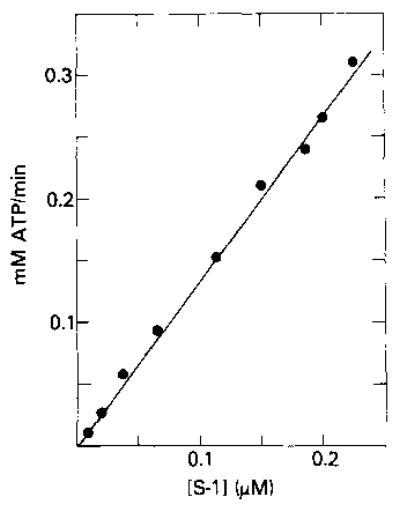
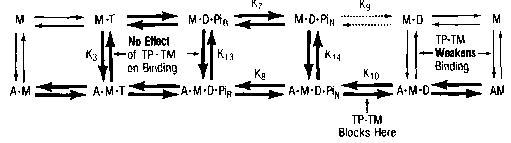
Relaxation of vertebrate skeletal muscle is thought to occur in the absence of Ca2+ as a result of tropomyosin physically blocking the binding of myosin to actin. This steric blocking model of muscle relaxation predicts that myosin subfragment 1 (S-1) will not bind to actin under conditions where the acto-S-1 ATPase rate is inhibited. Using stopped flow absorbance as a measure of binding, we have previously shown that when the rate of ATP hydrolysis is only 4% of the rate in the presence of Ca2+, S-1·ATP and S-1·ADP·Pi bind to actin-troponin-tropomyosin (regulated actin) with almost the same affinity as in the presence of Ca2+. This result has now been confirmed using sedimentation in an air-driven ultracentrifuge to directly measure the binding at pH 7.0, 25 °C, and μ = 18 mm. In the presence of Ca2+, the rate of ATP hydrolysis is more than 20 times greater than in the absence of Ca2+. In contrast, the association constant of S-1·ATP and S-1·ADP·Pi with regulated actin is virtually the same in the absence of Ca2+ (1.4 × 10⁴ m−1) as in the presence of Ca2+ (1.5 × 10⁴ m−1). Similarly, at 50 mm ionic strength, the ATPase rate is inhibited about 98% in the absence of Ca2+ although the association constant is not significantly changed compared to that in the presence of Ca2+. Finally, it has been shown that, at 18 mm ionic strength, the inhibition of the actin-activated ATPase rate in the absence of Ca2+ is due to a large decrease in the maximum ATPase rate (to 4% of the Ca2+ value) with only a small change in the apparent binding constant of S-1 to actin. These data do not support a simple steric blocking model of muscle relaxation. Rather they suggest that, in the absence of Ca2+, troponin-tropomyosin inhibits a kinetic step, perhaps Pi release, in the cycle of ATP hydrolysis.


Figures

Fig. 1
Fig. 1. Standard curve of the NH4+ EDTA ATPase assay used for determining the concentration of free S-1 in binding studies
Conditions: 5 mm ATP, 0.4 m NH4Cl, 35 mm EDTA, 25 mm Tris, pH 8.0, 25 °C.
Fig. 2
Fig. 2. Rate of ATP hydrolysis (A) and fraction of S-1 bound to regulated actin in the presence of ATP (B) as a function of the concentration of regulated actin at 18 mm ionic strength
Measurements were made in the presence (•) and absence (○) of Ca2+. In A, all rates were corrected for the rate of hydrolysis by S-1 (0.09 s−1) and the S-1 concentration was 0.1 or 1.5 μ m in the presence of Ca2+ and 0.27 or 2.7 μm in the absence of Ca2+. Conditions: 1 mm ATP, 3 mm MgCl2, 1mm EGTA (or 0.5 mm CaCl2), 10 mm imidazole, pH 7.0, 25 °C.
Fig. 3
Fig. 3. Double reciprocal plots of the fraction of S-1 bound, in the presence of ATP at 18 mm ionic strength, as a function of free actin concentration
Binding was measured using unregulated actin in the presence of Ca2+ (□), regulated actin in the presence of Ca2+ (•), and regulated actin in the absence of Ca2+ (○).
Fig. 4
Fig. 4. Rate of ATP hydrolysis by S-1 as a function of the concentration of regulated actin at 50 mm ionic strength in the presence (•) and absence (○) of Ca2+
All rates are corrected for the rate of hydrolysis by S-1 alone (0.1 s−1). The S-1 concentration was 0.2 μ m in the presence of Ca2+ and 2.0 μm in the absence of Ca2+. Conditions: 1 mm ATP, 3 mm MgCl2, 1 mm EGTA (or 0.5 mm CaCl2), 10 mm imidazole, 32 mm KC1, pH 7.0, 25 °C.
Fig. 5
Fig. 5. Double reciprocal plots of the fraction of S-1 bound, in the presence of ATP at 50 mm ionic strength, as a function of free actin concentration
Binding was measured with regulated actin in the presence (•) or absence (○) of Ca2+. Conditions: 2 mm ATP, 4 mm MgCl2,1 mm EGTA (or 0.5 mm CaCl2), 10 mm imidazole, 27 mm KC1, pH 7.0, 25°C.
Fig. 6
Fig. 6. Double reciprocal plots of S-1 ATPase against actin concentration at various ratios of native tropomyosin to actin at 18 mm ionic strength
Open symbols are in the presence of EGTA at molar ratios of native tropomyosin to actin of 0/7 (□), 1/7 (▿), 1.2/7 (▵), 1.5/7 (○), and 2/7 (⋄). For clarity, no line is drawn through the 0/7 ratio points. In the presence of Ca2+ (•), the ratio of tropomyosin to actin is 1.5/7. Conditions: 1 mm ATP, 3 mm MgCl2, 1 mm EGTA (or 0.5 mm CaCl2), 10 mm imidazole, pH 7.0, 25 °C.
Fig. 7
Fig. 7. Kinetic model of ATP hydrolysis by myosin
In this model, M is myosin or S-1, A is actin, T is ATP, D is ADP, TP·TM is troponin-tropomyosin, and the subscripts R and N are the refractory and nonrefractory states, respectively. Equilibrium constants involving formation of actin complexes are dissociation constants (e.g. K3 = [A][M·T]/[A·M·T]). For all other equilibrium constants, products are considered to be to the right of the reactants (e.g. K10 = [A·M·D][Pi]/(A·M·D·PiN]).

References

    1. Chalovich JM, Eisenberg E. Biophys J. 1981;33:232a. - PMC - PubMed
    1. Huxley AF, Niedergerke R. Nature (Lond) 1954;173:971–973. - PubMed
    1. Huxley HE, Hanson J. Nature (Lond) 1954;173:973–976. - PubMed
    1. Perry SV. Biochem Soc Trans. 1979;7:593–617. - PubMed
    1. Weber A, Murray JM. Physiol Rev. 1973;53:612–673. - PubMed