

Behavioral organization of reticular formation: studies in the unrestrained cat. I. Cells related to axial, limb, eye, and other movements
- PMID: 6619914
- PMCID: PMC8788630
- DOI: 10.1152/jn.1983.50.3.696
Behavioral organization of reticular formation: studies in the unrestrained cat. I. Cells related to axial, limb, eye, and other movements
Abstract
1. We have recorded single-unit activity in medial reticular formation (RF) units in unrestrained, behaving cats. A total of 306 cells have been analyzed. Neuronal activity was observed during a variety of natural, spontaneously occurring behaviors, during rapid eye movement (REM) and non-REM sleep states, after sensory stimulation, and during elicited reflexes.
2. Most RF units discharged maximally in conjunction with a specific movement or group of movements. The companion paper (41) deals with cells related to movements of the facial musculature, while the present paper deals with all other cell types.
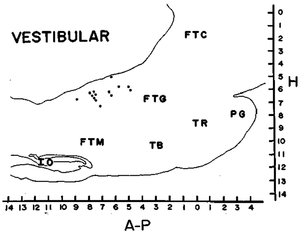
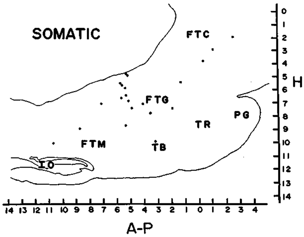
3. The most common RF cell types discharged during specific movements of the axial skeleton. Cells related to limb, respiratory, pharyngeal and laryngeal, jaw, and tongue movements were also observed. Reticular eye movement-related cells, previously investigated by others, were also seen in our unrestrained cats. A small percentage of cells were maximally activated by applied auditory, visual, vestibular, somatosensory, or proprioceptive stimuli.
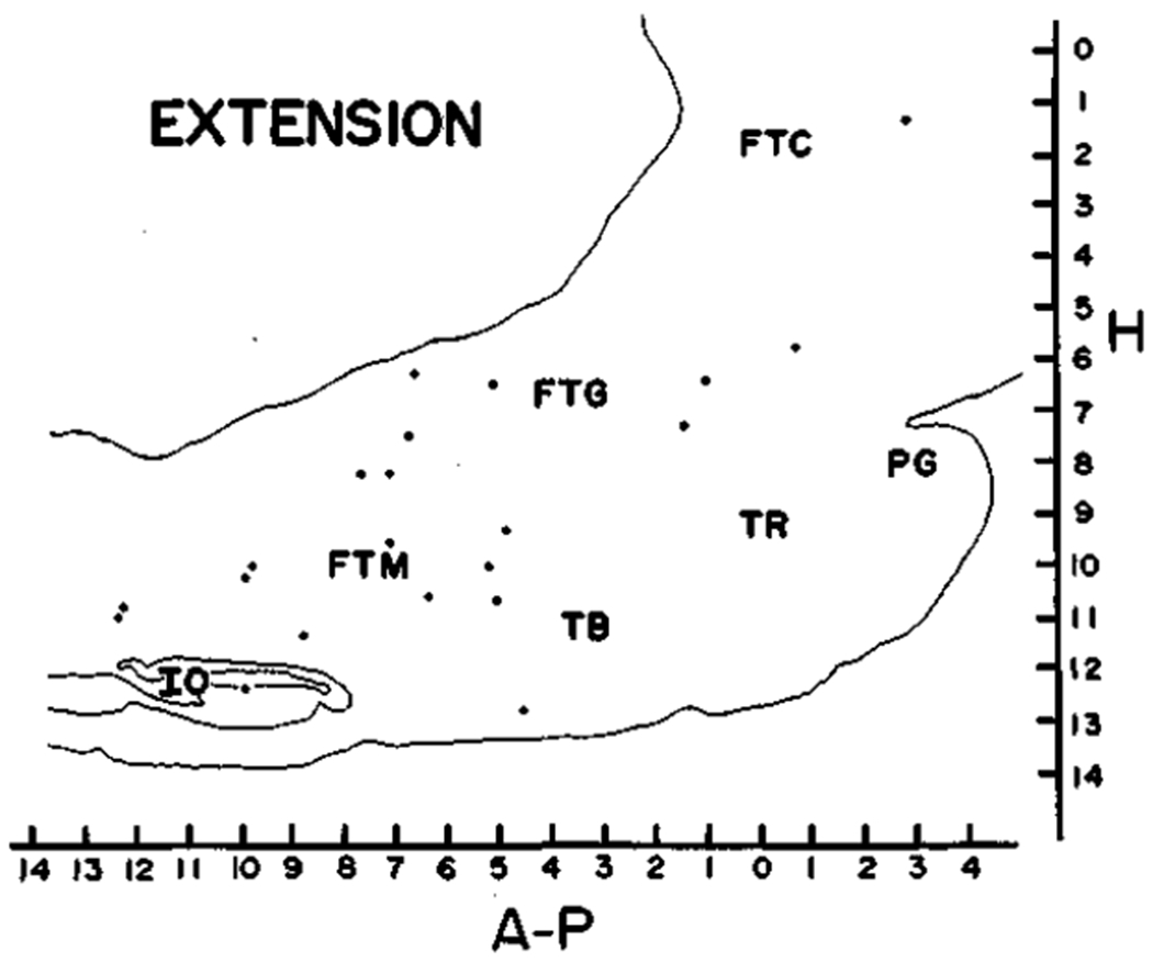
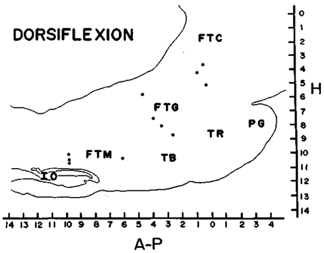
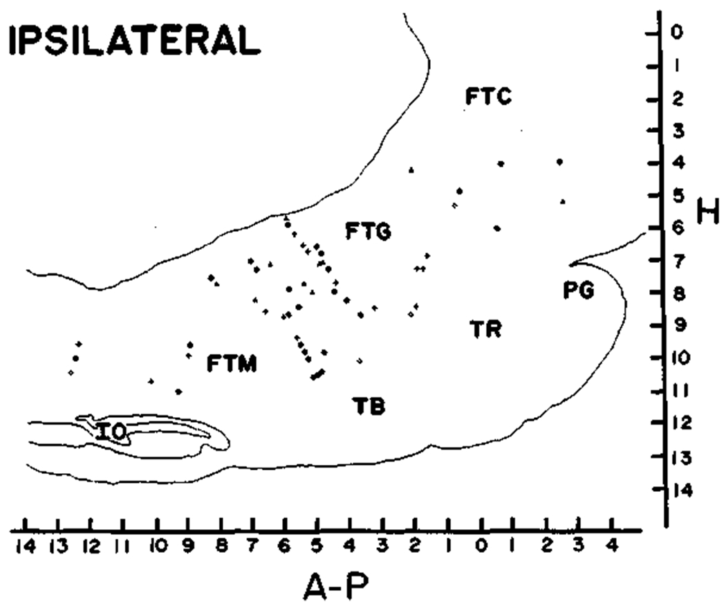
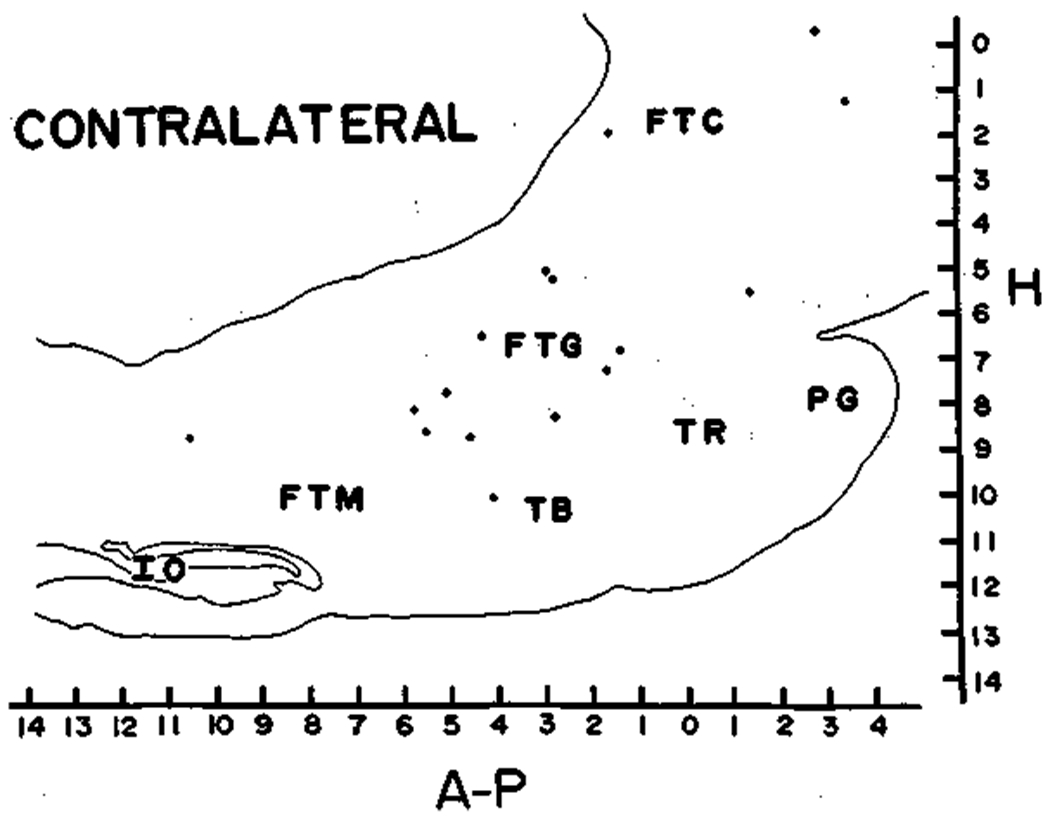
4. Cells related to axial movement, which as a group constituted 38.2% of all RF cells, could be subdivided into cells related to neck extension, neck dorsiflexion, and ipsilateral or contralateral movement of the spine.
5. Cells related to active ipsilateral movement, constituting 19.3% of all medial RF cells, were the single most common cell type in the RF. Thirty-four percent of these cells did not respond to passive head movement, while 48% responded to passive head movement to the contralateral side and 18% to passive movement to the ipsilateral side. Neck proprioceptors contribute to the passive movement response in certain of these cells.
6. Cells related to limb movement constituted 6.9% of the cells encountered. Most were related to movement of the proximal portion of one limb. Cells related to movement of the distal portion of the limb were quite rare, constituting only 1.0% of RF cells.
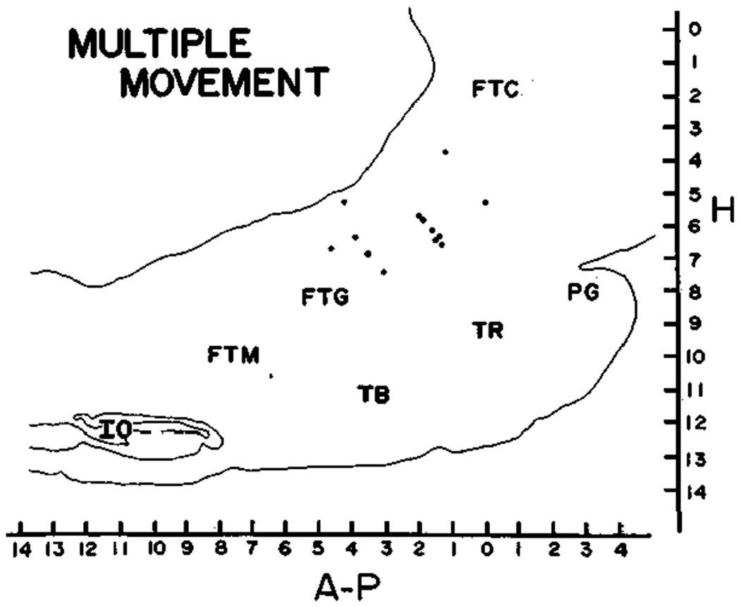
7. While most RF cells were active only in relation to a single, directionally specific movement, we found a cluster of pontine RF cells, which discharged in relation to several limb and neck movements.
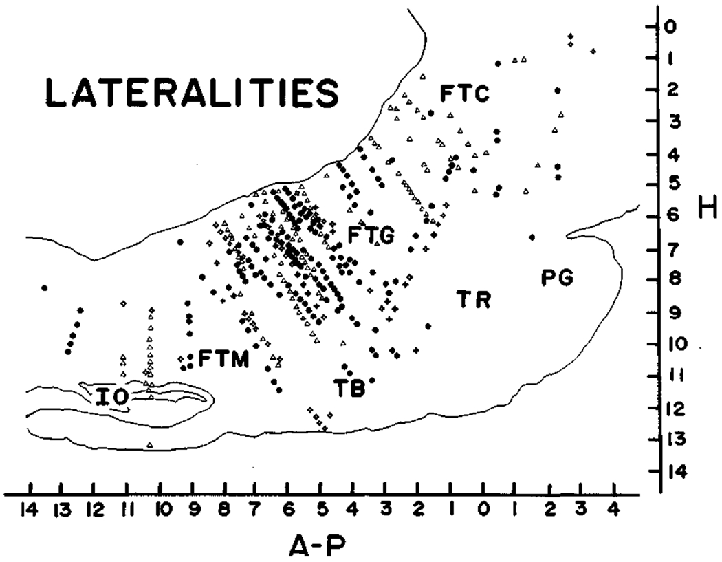
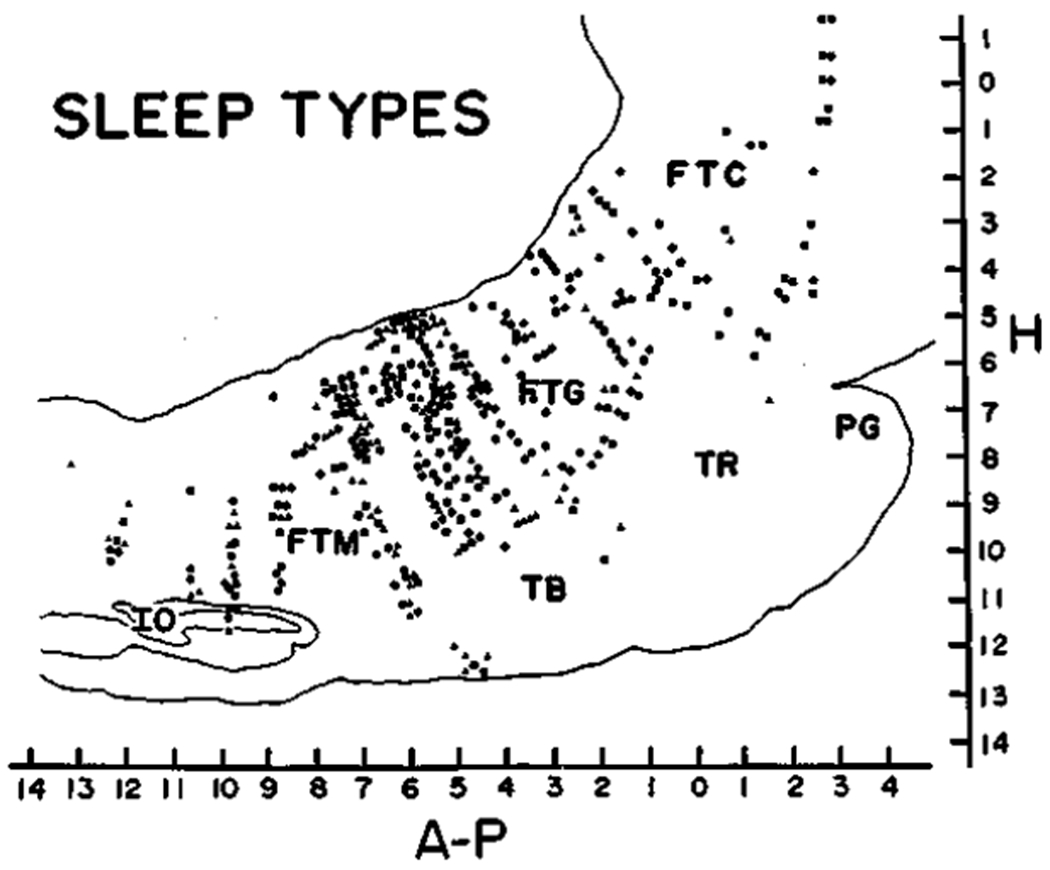
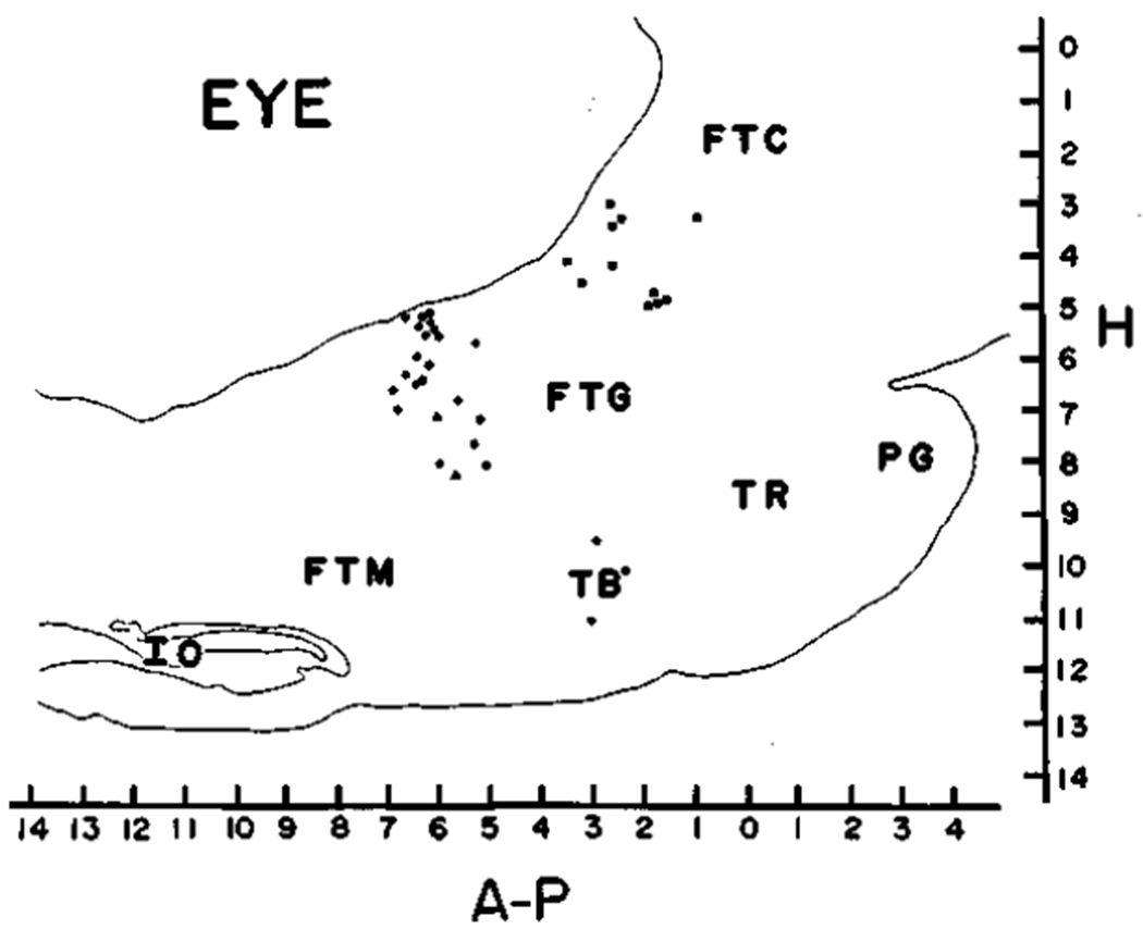
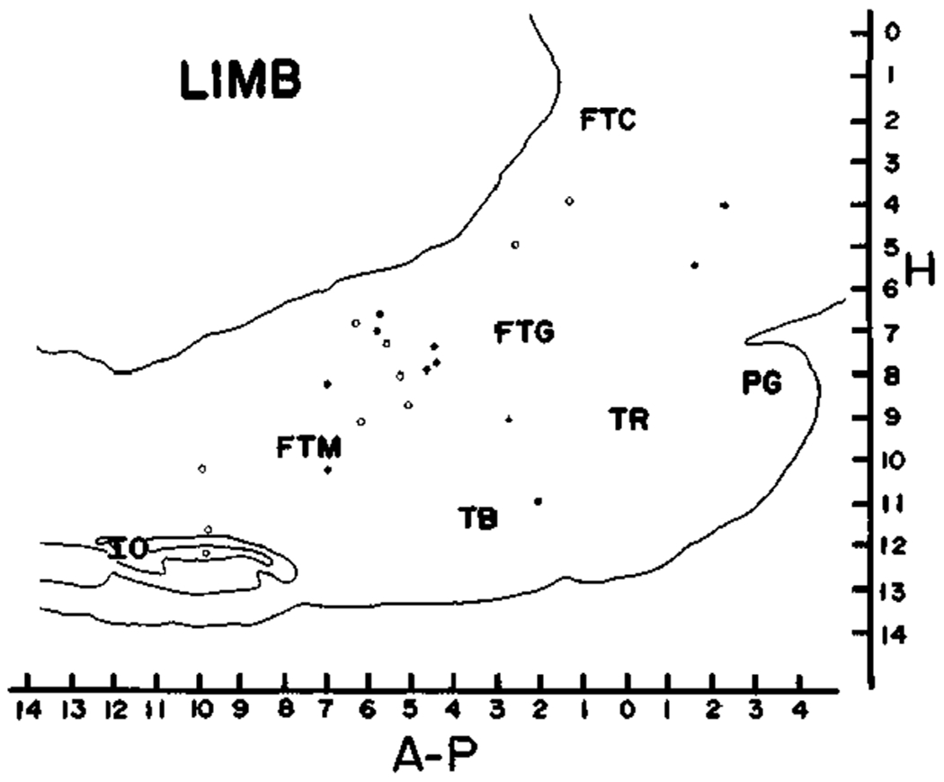
8. Cells related to several movements, eye movement, vestibular stimulation, and cells without spontaneous activity in sleep or waking were localized to restricted portions of the medial RF fields. Cells related to axial, proximal limb movements or somatic stimulation were intermingled throughout the entire region explored.
9. The unrestrained preparation allows the direct observation of the behavioral correlates of increased RF unit discharge. Most RF cells discharge in relation to a specific movement or group of movements of the axial musculature. Each movement-defined cell type has a different pattern of sleep, sensory and reflex activity, and anatomical localization. Anatomical intermingling of certain cell types may facilitate the synthesis of complex movement sequences from simpler elements commanded by individual RF cells.
Figures
Similar articles
-
Behavioral organization of reticular formation: studies in the unrestrained cat. II. Cells related to facial movements.J Neurophysiol. 1983 Sep;50(3):717-23. doi: 10.1152/jn.1983.50.3.717. J Neurophysiol. 1983. PMID: 6619915 Free PMC article.
-
Alterations in membrane potential and excitability of cat medial pontine reticular formation neurons during changes in naturally occurring sleep-wake states.Brain Res. 1984 Jan 30;292(1):169-75. doi: 10.1016/0006-8993(84)90903-x. Brain Res. 1984. PMID: 6320969
-
Reticulo-spinal neurons participating in the control of synergic eye and head movements during orienting in the cat. I. Behavioral properties.Exp Brain Res. 1987;66(2):339-54. doi: 10.1007/BF00243309. Exp Brain Res. 1987. PMID: 3595779
-
Studies of the role of the paramedian pontine reticular formation in the control of head-restrained and head-unrestrained gaze shifts.Ann N Y Acad Sci. 2002 Apr;956:85-98. doi: 10.1111/j.1749-6632.2002.tb02811.x. Ann N Y Acad Sci. 2002. PMID: 11960796 Review.
-
Head and eye movements in rats with pontine reticular lesions in comparison with primates: a scientific memoir and a fresh look at some old and 'new' data.Behav Brain Res. 2012 Jun 1;231(2):371-7. doi: 10.1016/j.bbr.2011.10.028. Epub 2011 Oct 25. Behav Brain Res. 2012. PMID: 22044476 Review.
Cited by
-
Clues to the functions of mammalian sleep.Nature. 2005 Oct 27;437(7063):1264-71. doi: 10.1038/nature04285. Nature. 2005. PMID: 16251951 Free PMC article. Review.
-
Monosynaptic excitatory connexions of reticulospinal neurones in the nucleus reticularis pontis caudalis with dorsal neck motoneurones in the cat.Exp Brain Res. 1990;80(2):277-89. doi: 10.1007/BF00228155. Exp Brain Res. 1990. PMID: 2358043
-
Is there a brainstem substrate for action selection?Philos Trans R Soc Lond B Biol Sci. 2007 Sep 29;362(1485):1627-39. doi: 10.1098/rstb.2007.2057. Philos Trans R Soc Lond B Biol Sci. 2007. PMID: 17428776 Free PMC article.
-
The echidna Tachyglossus aculeatus combines REM and non-REM aspects in a single sleep state: implications for the evolution of sleep.J Neurosci. 1996 May 15;16(10):3500-6. doi: 10.1523/JNEUROSCI.16-10-03500.1996. J Neurosci. 1996. PMID: 8627382 Free PMC article.
-
The Vestibular-Evoked Postural Response of Adolescents with Idiopathic Scoliosis Is Altered.PLoS One. 2015 Nov 18;10(11):e0143124. doi: 10.1371/journal.pone.0143124. eCollection 2015. PLoS One. 2015. PMID: 26580068 Free PMC article.
References
-
- AMASSIAN VE AND DEVITO RV Unit activity in reticular formation and nearby structures. J. Neurophysiol 17: 575–603, 1954. - PubMed
-
- BACH-Y-RITA P Convergent and long-latency unit responses in the reticular formation of the cat. Exp. Neurol 9: 327–344, 1964. - PubMed
-
- BERMAN AL The Brain Stem of the Cat. Madison: University of Wisconsin, 1968.
Publication types
MeSH terms
Grants and funding
LinkOut - more resources
Full Text Sources
Miscellaneous