. 1984 May;309(5964):155-7.
doi: 10.1038/309155a0.

Regional specialization of retinal glial cell membrane

Regional specialization of retinal glial cell membrane

E A Newman. Nature. 1984 May.

Abstract

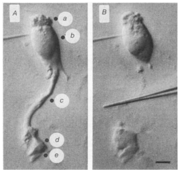
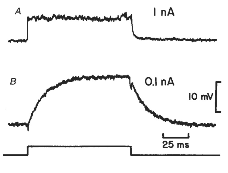
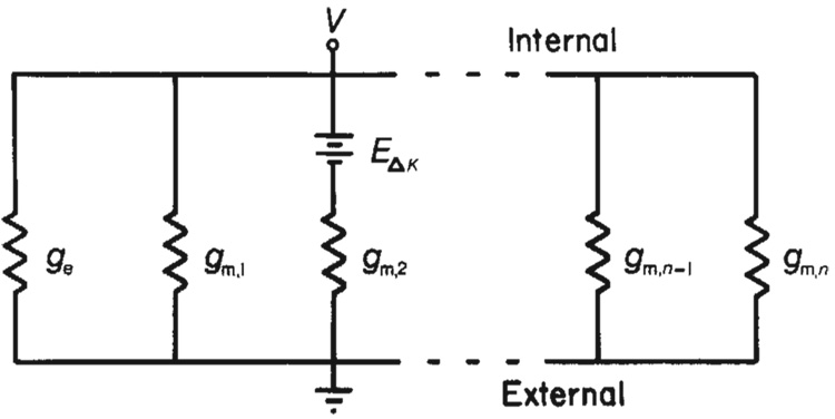
Neural activity generates increases in extracellular K+ concentration, [K+]0, which must be regulated in order to maintain normal brain function. Glial cells are thought to play an important part in this regulation through the process of K+ spatial buffering: K+-mediated current flow through glial cells redistributes extracellular K+ following localized [K+]0 increases. As is the case in other glia, the retinal Müller cell is permeable almost exclusively to K+ . Recent experiments have suggested that this K+ conductance may not be distributed uniformly over the cell surface. In the present study, two novel techniques have been used to assess the Müller cell K+ conductance distribution. The results demonstrate that 94% of all membrane conductance lies in the endfoot process of the cell. This strikingly asymmetric distribution has important consequences for theories concerning K+ buffering and should help to explain the generation of the electroretinogram.


Figures

Fig. 1
Fig. 1. Photomicrographs of a dissociated salamander Müiller cell viewed with Nomarski optics
A, Intact cell penetrated in the nuclear region. Solid circles indicate locations of the K+-ejection pipette used in K+-ejection experiments. a, Distal end; b, nuclear region; c, stalk; d, lateral face of endfoot; e, proximal face of endfoot. B, Same cell after stalk has been cut by glass needle (shown in the centre of the photograph). Scale bar, 10 µm. Dissociated cells were prepared by incubating isolated retinae in Ringer’s solution containing papain (Sigma P-3125; 0.75 mg ml−1) for 30 min followed by gentle mechanical separation, as described previously. The Ringer’s contained (in mM): 82.5 NaCl, 27.5 NaHCO3, 2.5 KC1, 1.8 CaCl2, 1.0 MgCl2, 10.0 dextrose, equilibrated with 5 % CO2 in O2. Dissociated cells adhered to the bottom of the recording chamber, a glass slide coated with gelatin and concanavalin A (Sigma C-2010).
Fig. 2
Fig. 2. Müller cell depolarizations evoked by current pulses applied through an intracellular pipette in the nuclear region of the cell
A, Response from an intact cell. Resistance = 7.4 MΩ; τ = 0.86 ms (1 nA current pulse). B, Response from the same cell after the endfoot process was severed. Resistance = 156 MΩ; τ = 16.7 ms (0.1 nA current pulse). The time course of the current pulse is indicated at the bottom.
Fig. 3
Fig. 3. Responses of a dissociated Müller cell (penetrated near the nucleus) to K+ ejections from an extracellular pipette
The labels correspond to ejection locations, shown in Fig. 1A. Each trace is an average of eight sweeps. Onset and duration of a 5-ms pressure pulse is indicated at the bottom. Traces a–c are expanded vertically fivefold relative to traces d and e.
Fig. 4
Fig. 4. Ohmic model of Müller cell simulates K+ ejection results
The idealized cell is divided into n membrane segments each having conductance gm and one membrane segment having conductance ge, representing the high conductance endfoot. If we assume that a K+ ejection depolarizes a single membrane segment, the amplitude of a K+ ejection response can be calculated by interposing a voltage source in series with that membrane segment. This depolarization is represented by EΔK in the model. If the endfoot membrane segment is depolarized, the entire cell response, Ve, will equal EΔK ge/(ngm + ge). If a membrane segment in some other cell region is depolarized, the cell response, Vm, will equal EΔKgm/(ngm + ge). The ratio of responses, Ve/Vm, equals ge/gm. If more than one membrane segment is depolarized by a K+ ejection, the ratio of responses will be reduced. If five segments are depolarized equally by a K+ ejection, for example, the ratio of responses (endfoot to low conductance region) equals (ge + 4gm)/5gm, or roughly 1/5 of ge/gm when gegm. During experimental K+ ejections directed towards the endfoot, an area significantly larger than the endfoot region was depolarized (unpublished observations). Consequently, the amplitude ratios of the responses shown in Fig. 3 lead to an underestimation of the value of the specific membrane conductance of the endfoot compared with the conductance of other cell regions. This analysis does not incorporate the effects of internal cell resistance on K+ responses. Internal resistance will, in fact, reduce the amplitude of the endfoot K+ response relative to the amplitudes of responses of cell segments nearer the recording site.

References

    1. Gardner-Medwin AR. J. exp. Biol. 1981;95:111–127. - PubMed
    1. Orkand RK, Nicholls JG, Kuffler SW. J. Neurophysiol. 1966;29:788–806. - PubMed
    1. Trachtenberg MC, Pollen DA. Science. 1969;167:1248–1252. - PubMed
    1. Gardner-Medwin AR. J. Physiol, Lond. 1983;335:393–426. - PMC - PubMed
    1. Newman EA. Soc. Neurosci. abstr. 1981;7:275.

Publication types