

doi: 10.1038/300611a0.
Dramatic growth of mice that develop from eggs microinjected with metallothionein-growth hormone fusion genes
- PMID: 6958982
- PMCID: PMC4881848
- DOI: 10.1038/300611a0
Item in Clipboard
Dramatic growth of mice that develop from eggs microinjected with metallothionein-growth hormone fusion genes
Nature.
.
Abstract
A DNA fragment containing the promoter of the mouse metallothionein-I gene fused to the structural gene of rat growth hormone was microinjected into the pronuclei of fertilized mouse eggs. Of 21 mice that developed from these eggs, seven carried the fusion gene and six of these grew significantly larger than their littermates. Several of these transgenic mice had extraordinarily high levels of the fusion mRNA in their liver and growth hormone in their serum. This approach has implications for studying the biological effects of growth hormone, as a way to accelerate animal growth, as a model for gigantism, as a means of correcting genetic disease, and as a method of farming valuable gene products.
Figures
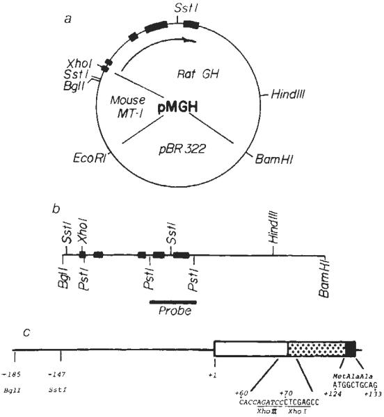
Construction of the fusion plasmid pMGH. a, The unique BglII site of the MT-I genomic clone was converted to a XhoI site by digesting with BglII, followed by filling in the sticky ends with Klenow fragment of DNA polymerase in the presence of all four dNTPs; then XhoI linkers (CCTCGAGG) were ligated to the blunt ends and bacterial strain RR1 was transformed with this DNA. The PvuII site of pBR322 was converted to a BamHI site by a similar procedure, then the 4.1-kb XhoI–BamHI fragment containing the MT-I promoter and pBR was ligated to a 4.8-kb XhoI–BamHI fragment containing the rat GH structural gene (see refs 10, 30) to give pMGH (8.9 kb). b, The fragment used for injection was a 5.0-kb BglI–BamHI fragment which was isolated from an agarose gel by the NaClO4 method. For genomic Southern blots, a 1.0-kb PstI fragment spanning exons 4 and 5 was isolated and nick-translated and used as a hybridization probe. c, The predicted structure of exon 1 of the fusion gene. The line and open box represent MT-1 untranslated sequences, the stippled box represents GH untranslated sequences and the solid box represents the beginning of the GH coding region (see refs 28 and for complete sequence information).
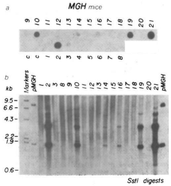
Analysis of DNA from transgenic mice for MGH sequences. a, DNA dot hybridization was used to detect mice with MGH sequences and quantitate their abundance. A nick-translated PstI probe (see Fig. 1b) was used (6 h exposure). Numbers correspond to the MGH mice examined; c, controls, b, DNA (5 μg) from MGH mice was restricted with SstI electrophoresed on a 1% agarose gel, transferred to nitrocellulose and hybridized with the nick-translated PstI probe shown in Fig. 1b. pMGH (13 pg, left and 130 pg, right) was included as a hybridization standard. Markers are HindIII-cut λ DNA end-labelled with 32P.
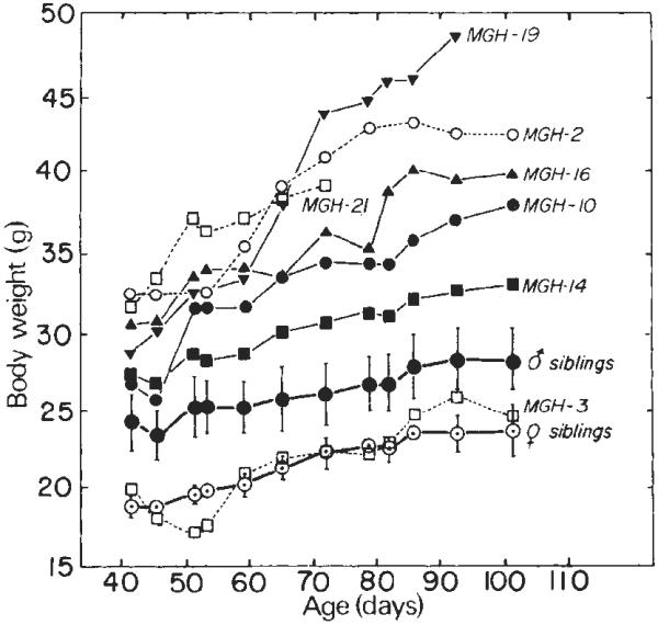
Growth of MGH mice. Microinjected eggs (SJL × C57) were transferred to oviducts of foster mothers on 14 April; 21 animals were born 3 weeks later. At 33 days old they were weaned and the drinking water was supplemented with 76 mM ZnSO4. The body weights of the males are shown as solid symbols; the mean weight (± s.d.) of 11 siblings not containing MGH sequences is also shown. The female weights are represented by open symbols; means (± s.d.) of three siblings are indicated also. MGH-21 died on day 72. A partial hepatectomy was performed on MGH-19 on day 56 and it was taken off Zn thereafter; it was killed on day 100. All mice were taken off Zn after day 83.
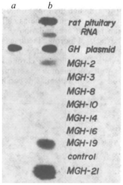
Slot-blot analysis of liver RNAs from transgenic mice. Mouse liver RNA was purified by homogenizing a liver slice in 1 × SET (1% SDS, 5 mM EDTA, 10 mM Tris pH 7.5), 200 μg ml−1 proteinase K (Boehringer) followed by phenol/chloroform extraction, ethanol precipitation, DNase I treatment (Worthington; DPFF), a second phenol/chloroform extraction, then re-precipitation with ethanol and finally resuspension for A260 determination. Liver RNA (30 μg) was diluted to 100 μl in 2X SSC. Samples were brought to 0.5 M NaOH or 0.5 M NaCl and incubated at 65 °C for 30 min. 140 μl of 20 × SSC and 160 μl of 12.3 M formaldehyde were added, incubated at 65 °C for another 15 min and applied to a nitrocellulose sheet resoaked in 20 × SSC through a slot-blot device. The nitrocellulose was baked at 80 °C for 2 h, prehybridized for 5 h and then hybridized with nick-translated 12P-rGH cDNA (5 × 10⁶ c.p.m. in 4 ml at 42 °C overnight), washed in 0.1 × SSC at 42 °C, and exposed to X-ray film at −70 °C for 24 h. a, Samples were treated with 0.5 M NaOH and neutralized with HCl. b, Samples were incubated with an equivalent amount of NaCl. 40 and 20 ng of rat pituitary poly(A)+ RNA in the presence of 30 μg of control liver RNA were applied to slots as GH mRNA controls. 1 ng of rGH gene plasmid, also in the presence of 30 μg of control liver RNA, was applied as a base-resistant control.
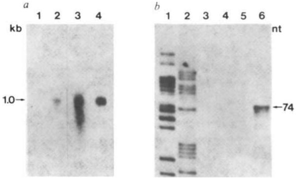
Structure of MGH mRNAs in liver of transgenic mice. a, Northern blot analysis. RNAs were resuspended 10 mM NaH2PO4, pH 7.4/50% formamide/2.2 M formaldehyde, heated at 65 °C for 5 min, and subjected to electrophoresis on a slab gel composed of 1.5% agarose in 10 mM NaH2PO4, pH 7.4/0.55 mM EDTA/1.1 M formaldehyde. The running buffer was 10 mM NaH2PO4, pH 7.4/0.5 M formaldehyde. The gel was stained with acridine orange to identify rRNA markers, photographed, incubated for 90 min with 50 mM NaOH, and then neutralized with two washes of 0.2 M NaOAc, pH 4.3. The RNA was then transferred to diazotized paper and prehybridized as described elsewhere. Lane 1, total liver RNA (15 μg) from a control littermate; lane 2, total liver RNA (15 μg) from MGH-21; 3, total mouse pituitary RNA; 4, 40 ng of rat pituitary mRNA, poly(A)+. Lanes 1 and 2 exposed for 48 h; lanes 3 and 4 exposed for 5 h. b, Single-strand-specific nuclease protection assay. 20 μg of RNA were hybridized at 47 °C for 5 h in 40 μl of a solution containing 40 mM PIPES pH 6.4, 0.4 M NaCl, 80% formamide and 50,000 c.p.m. gel-purified 221-bp SstI–XhoI fragment of pMGH end-labelled at the XhoI site with 32P (see Fig. 1c). The samples were then diluted with 0.3 ml of 280 mM NaCl, 30 mM NaOAc pH 4.4, 4.5 mM ZnSO4, 20 μg ml−1 salmon sperm DNA and 150 units of mung bean single-strand-specific nuclease (Collaborative Research) and incubated at 47 °C for 1 h. Samples were ethanol-precipitated, resuspended in 90% formamide containing bromophenol blue and Xylene Cyanol FF, loaded on to an 8% acrylamide–urea sequencing gel, electrophoresed for 1.5 h at 2,000 V, dried and autoradiographed for 7 days at −70 °C with an intensifying screen. Lanes 1 and 2, sequencing ladder used for size standards; 3, MGH-3 RNA; 4, control liver RNA; 5, mouse pituitary RNA; 6, MGH-21 RNA.
References
Publication types
MeSH terms
Substances
Grants and funding
LinkOut - more resources
Full Text Sources
Other Literature Sources
Molecular Biology Databases
