. 1980 Dec;62(8):1362-9.

Anatomy of the microvasculature of the tibial diaphysis of the adult dog

Anatomy of the microvasculature of the tibial diaphysis of the adult dog

J A Lopez-Curto et al. J Bone Joint Surg Am. 1980 Dec.

Abstract

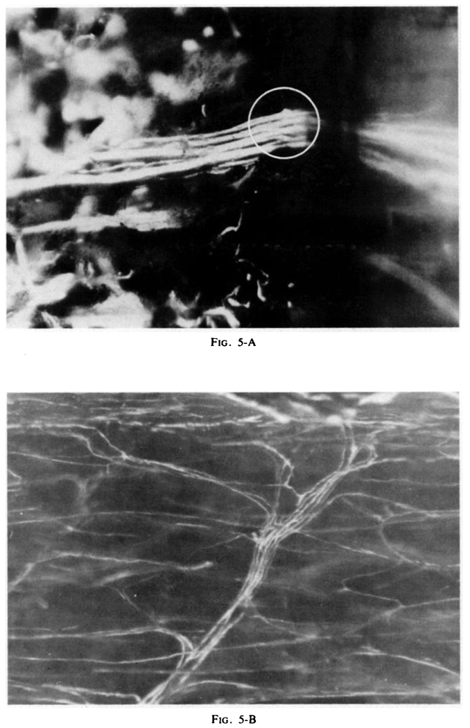
The microvasculature in the cortex and marrow of the adult canine tibial diaphysis was filled with the silicone elastomer Microfil, the bone was decalcified, and the water was replaced with methylsalicylate to permit three-dimensional visualization of the microvascular arrangements. The tibial nutrient artery was seen to supply the marrow and the cortex via parallel, independent sets of arterioles and terminal capillary beds. No arteriolar or capillary anastomoses were observed linking these separate beds. The major portion of the venous drainage was found to be via small venules through the cortex into periosteal veins. Many small venules draining the medullary capillaries penetrated the cortex, and there were a few larger emissary veins, including the nutrient vein. Because the marrow and cortex have separate capillary beds in parallel, microsphere deposition should be appropriate for estimating the regional blood flows.

Clinical relevance: The results of this study should be of concern to surgeons who perform whole diaphyseal bone replacements, as the effluent venous vessels are important in re-establishing the circulation by microsurgical methods.


Figures

FIG. 1
FIG. 1
Diagram of circulation in the tibial diaphysis.
FIG. 2
FIG. 2
Composite photograph at low magnification of transversely cut fragment of tibia infused with Microfil . Note the rich vascularity of the medullary cavity and the relative sparseness of the cortical vascularity. The complex at the endosteum on the right is the tibial nutrient artery and vein. The periosteum has been removed (approximately × 20).
FIG. 3
FIG. 3
Transverse section of the tibial diaphysis. The dividing branch of the nutrient artery can be seen traversing the medullary cavity among sinusoids and penetrating cortex . Branches undergo further subdivision in the cortex and anastomose with periosteal vessels . This photograph was obtained by placing the specimen at an angle of 30 degrees, which allowed visualization of the inner surface of the periosteum (approximately × 60).
FIG. 4
FIG. 4
A branch of the nutrient artery penetrates the cortex and undergoes multiple subdivisions into even smaller arterioles. In the upper right-hand corner (box), an intracortical pattern of multiple subdivisions can be seen. The pattern of drainage into periosteal veins also can be appreciated (approximately × 60).
FIG. 5
FIG. 5
The circle indicates the single orifice of the entry of the artery into cortex in the endosteal surface. Note that before entering the orifice the artery has divided into multiple branches (approximately × 180). Detail of Fig. 4 , showing the intracortical pattern of these divisions within the osteal canal( approximately × 200).
FIG. 6
FIG. 6
A nutrient-artery branch can be seen subdividing in the midst of the sinusoidal network at the upper left-hand corner. In the area marked with arrows, marrow structures were partially dissected to show detail. A minute branch can be seen entering a sinusoid. The artery then subdivides, as illustrated in Fig. 5-A, but the uppermost branch, instead of entering the cortex, separates itself from the main bundle and, running in the endosteal surface, supplies the sinusoids (arrows) (approximately × 180).
FIG. 7
FIG. 7
Sinusoidal vein and transcortical so-called emissary vein. The large vein in the center (approximately 100 micrometers in diameter as it enters the cortex) drains blood from the marrow (below)(approximately × 50).
FIG. 8
FIG. 8
Composite photograph illustrating the formation of the collecting sinusoid at the endosteal surface and its connection with a vein within the cortex. The vein and artery are in an osteal canal (approximately × 50).
FIG. 9
FIG. 9
Branching of an arteriole (a) within the cortex; the venae comitantes (v) here have an arrangement common to many other tissues, draining the cognate capillary bed of the arteriole in two directions, to the periosteum (above) or near its source in the medulla (below) and thence to an emissary vein such as is shown in Fig. 7. This type of arrangement appears to contrast with the arrangement suggested by Fig. 2, which is at low power, but fits that seen in Figs. 3, 4, 5-A, and 5-B, which are at higher power (approximately × 120).
FIG. 10
FIG. 10
Venous network on the surface of the periosteum of the cortical bone (approximately × 40).

References

    1. Brålnemark P-I. Vital Microscopy of Bone Marrow in Rabbit. Scandinavian J. Clin. and Lab. Invest. 1959;38 Supplement:5–82. - PubMed
    1. Cofield RH, Bassingthwaighte JB, Kelly PJ. Strontium-85 Extraction During Transcapillary Passage in Tibial Bone. J. Appl. Physiol. 1975;39:596–602. - PMC - PubMed
    1. De Bruyn PPH, Breen PC, Thomas TB. The Microcirculation of the Bone Marrow. Anat. Rec. 1970;168:55–68. - PubMed
    1. Heymann MA, Payne BD, Hoffman JIE, Rudolph AM. Blood Flow Measurements with Radionuclide-Labeled Particles. Prog. Cardiovasc. Dis. 1977;20:55–79. - PubMed
    1. Kelly PJ. Comparison of Marrow and Cortical Bone Blood Flow By 125I-Labeled 4-Iodoantipyrine(I-Ap) Washout. J. Lab. and Clin. Med. 1973;81:497–505. - PubMed

Publication types