. 1993 Oct 15;336(3):321-30.
doi: 10.1002/cne.903360302.

Glutamatergic and cholinergic projections to the pontine inhibitory area identified with horseradish peroxidase retrograde transport and immunohistochemistry

Affiliations

Glutamatergic and cholinergic projections to the pontine inhibitory area identified with horseradish peroxidase retrograde transport and immunohistochemistry

Y Y Lai et al. J Comp Neurol. .

Abstract

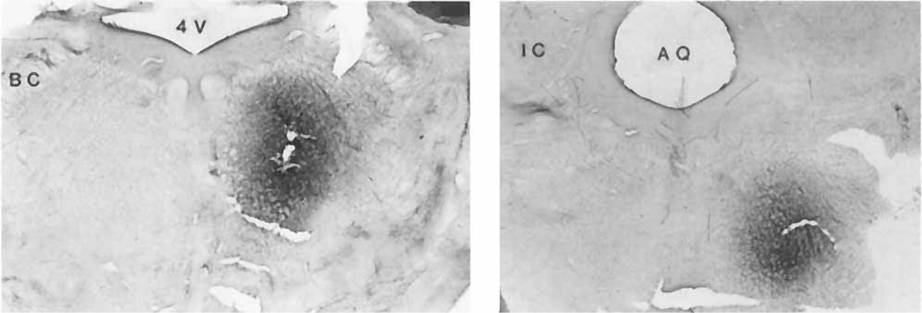
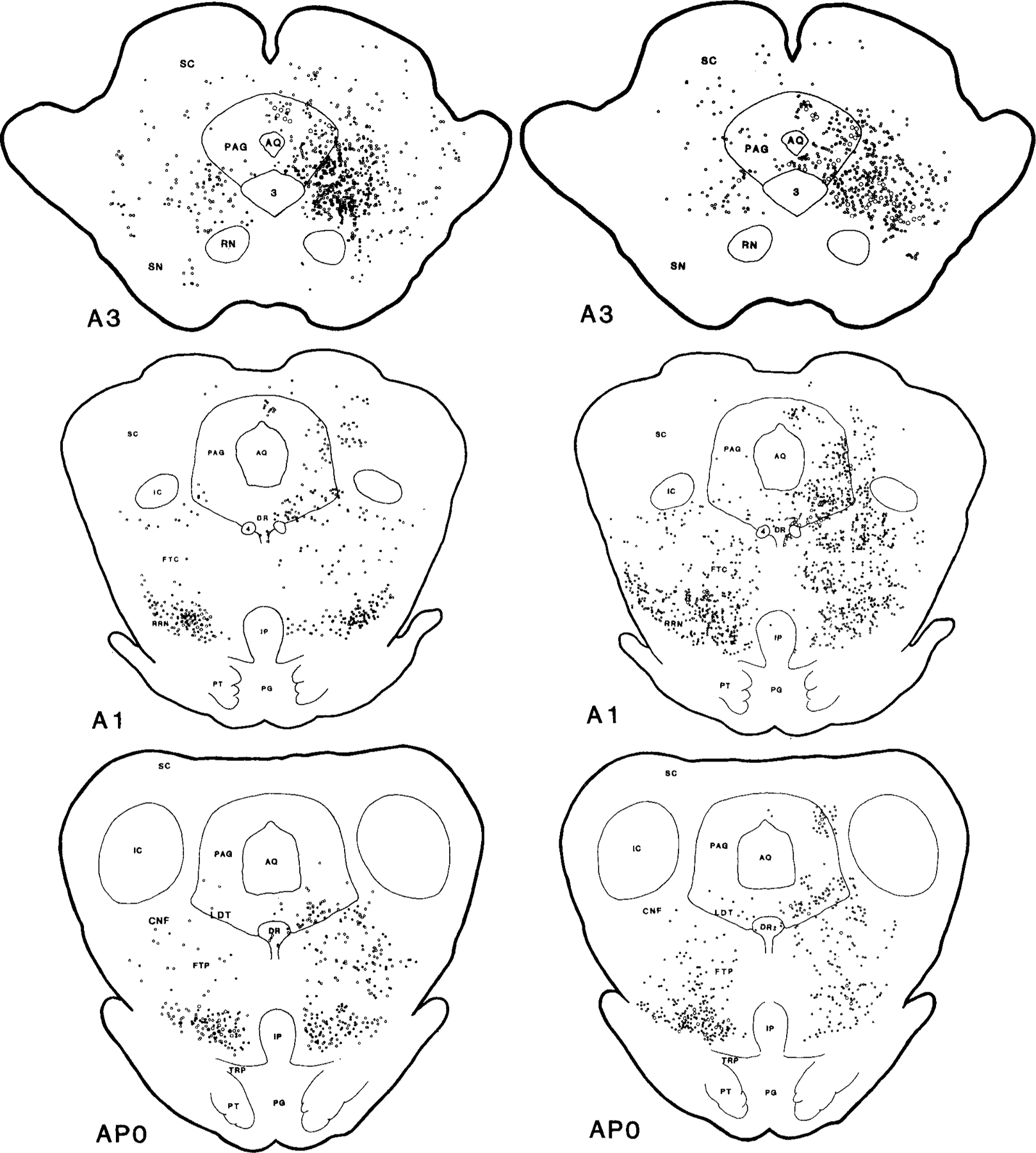
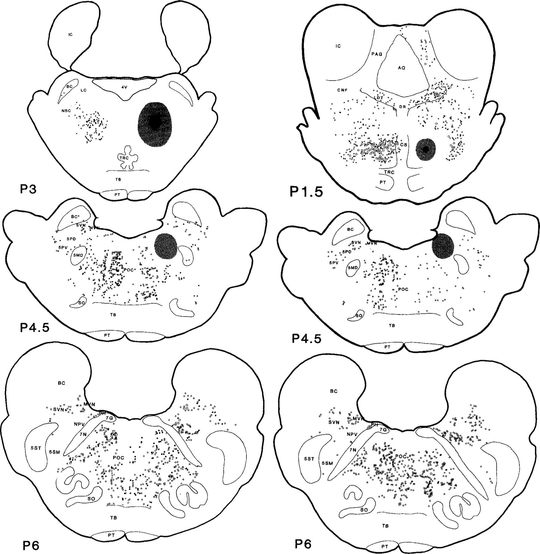
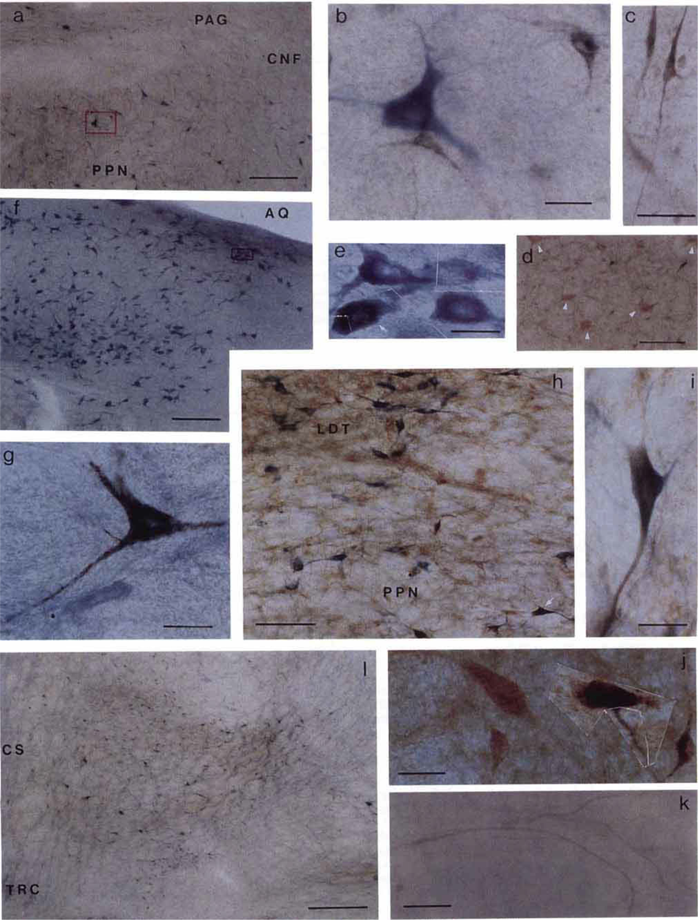
Previous studies in our laboratory have shown that microinjection of acetylcholine and non-N-methyl-D-aspartate (NMDA) glutamate agonists into the pontine inhibitory area (PIA) induce muscle atonia. The present experiment was designed to identify the PIA afferents that could be responsible for these effects, by use of retrograde transport of wheat germ agglutinin conjugated horseradish peroxidase (WGA-HRP), glutamate immunohistochemistry and NADPH-diaphorase staining techniques. Experiments were performed in both decerebrate and intact cats. Dense retrograde WGA-HRP labelling was found in neurons in the periaqueductal gray (PAG) and mesencephalic reticular formation (MRF) at the red nucleus (RN) level, ventral portion of paralemniscal tegmental field (vFTP), retrorubral nucleus (RRN), contralateral side of PIA (cPIA), pontis reticularis centralis caudalis (PoC), and most rostral portion of the nucleus parvicellularis (NPV) and nucleus praepositus hypoglossi (PH) at the level of the pontomedullary junction; moderate labelling was seen in pedunculopontine nucleus, pars compacta (PPNc), laterodorsal tegmental nucleus (LDT), superior colliculus (SC), MRF and PAG at the level caudal to RN, medial and superior vestibular nuclei, and principle sensory trigeminal nucleus (5P); and light labelling was seen in dorsal raphe (DR) and locus coeruleus complex (LCC). The projection neurons were predominantly ipsilateral to the injection site, except for both vFTP and RRN, which had more projection cells on the contralateral side. Double labelled WGA-HRP/NADPH-d neurons could be found in PPNc and LDT. Double labelled WGA-HRP/glutamatergic neurons could be seen at high densities in MRF, RRN, vFTP, and cPIA, moderate densities in SC, LDT, PPNc, PoC, and NPV, and low densities in PH, 5P, DR, LCC, and PAG.(ABSTRACT TRUNCATED AT 250 WORDS)


Figures

Fig. 1.
Fig. 1.
Photographs show two examples of injection sites. Magnification: × 2.5.
Figs. 2 and 3.
Figs. 2 and 3.
Serial coronal sections of the mesopontine brainstem illustrate the wheat germ agglutinin conjugated horseradish peroxidase (WGA-HRP) labelled projecting neurons also containing NADPH-d (right) and glutamate (left). Dots represent WGA-HRP neurons double labelled with either NADPH-d or glutamate. Circles represent neurons labelled with WGA-HRP only. The small and large symbols represent one and five neurons, respectively. Each view is based on a 50 µm section from one cat. The black and stippled areas represent the WGA-HRP injection center and diffusion areas, respectively. The histology was reconstructed and the rostrocaudal levels were modified according to Berman’s atlas (1968). A, anterior; P, posterior to stereotaxic zero.
Figs. 2 and 3.
Figs. 2 and 3.
Serial coronal sections of the mesopontine brainstem illustrate the wheat germ agglutinin conjugated horseradish peroxidase (WGA-HRP) labelled projecting neurons also containing NADPH-d (right) and glutamate (left). Dots represent WGA-HRP neurons double labelled with either NADPH-d or glutamate. Circles represent neurons labelled with WGA-HRP only. The small and large symbols represent one and five neurons, respectively. Each view is based on a 50 µm section from one cat. The black and stippled areas represent the WGA-HRP injection center and diffusion areas, respectively. The histology was reconstructed and the rostrocaudal levels were modified according to Berman’s atlas (1968). A, anterior; P, posterior to stereotaxic zero.
Fig. 4.
Fig. 4.
Photomicrographs showing labelled neurons in the brain­stem. Sections shown in a, b, e, f, g, k, and l were processed with tetramethylbenzidine (TMB) and NADPH-d histochemical staining; sections c, d, and j were processed with TMB and glutamate immunohistochemical staining; and sections h and i were stained with TMB, NADPH-d, and glutamate immunohistochemical technique. The areas within the red boxes in a and f are shown at higher magnification in b and e, respectively. a: Low power photomicrograph shows the NADPH-d positive (blue) and WGA-HRP (black) neurons in PPN. b: Large single labelled NADPH-d positive cholinergic neuron had multiple neuronal processes. Two crystallized WGA-HRP neurons had a relatively smaller size compared with the NADPH-d positive neuron. c: Photomicrograph showing two small spindle shaped cells with elongated neuronal processes in RRN. d: WGA-HRP negative but glutamate-like immunoreactive giant neurons (white arrowheads) in cPIA. e: High power photomicrograph montage shows the NADPH-d positive (blue) and double labelled NADPH-d positive and WGA-HRP (white arrow) neurons in LDT taken from inset in f. The black crystal of TMB products was mixed in the homogenous blue NADPH-d positive reaction in the double labelled neuron. g: Large single labelled WGA-HRP neuron taken from the POC. h, i: Double labelled glutamate (brown) and NADPH d (blue) immunohistochemical stained neuron taken from the PPN (h, white arrow). j: Montage of two single labelled glutamate-like reactive neurons and two double labelled glutamate and WGA-HRP neurons seen in contralateral side of the PIA. k: TMB stained crossing fibers from the contralateral side of the PIA projecting to the injection site were seen in the CS area. l: Low power photomicrograph showing high density of the projection neurons in ventral part of the FTP. See text for details. Scale bars: a, d, and f = 200 µm; b, e, g, i, and j = 20 µm; c and k = 50 µm; h = 100 µm; 1 = 500 µm.

References

    1. Beitz AJ, Larson AA, Monaghan P, Altschuler RA, Mullet MM, and Madl JE (1986) Immunohistochemical localization of glutamate, glutaminase, and aspartate aminotransferase in neurons of the pontine nuclei of the rat. Neuroscience 17:741–753. - PubMed
    1. Berman AL (1968) The Brain Stem of the Cat Madison: University of Wisconsin Press.
    1. Fonnum F (1991) Neurochemical studies on glutamate-mediated neuro-transmission. In Meldrum BS, Moroni F, Simon RP, and Woods JH (eds): Excitatory Amino Acids New York: Raven Press, pp. 15–25.
    1. George R, Haslett WL, and Jenden DJ (1964) A cholinergic mechanism in the brainstem reticular formation: Induction of paradoxical sleep. Int. J. Neuropharmacol 3:541–552. - PubMed
    1. Hepler JR, Petrusz P, and Rustioni A (1986) Antisera to GABA, glutamate and aspartate: Characterization by immunoabsorption and immunocytochemistry. J. Histochem. Cytochem 34:110. - PubMed

Publication types

LinkOut - more resources