. 1993;3(2):201-13.

A cluster of five nuclear proteins regulates keratin gene transcription

Affiliations

A cluster of five nuclear proteins regulates keratin gene transcription

M Ohtsuki et al. Gene Expr. 1993.

Abstract

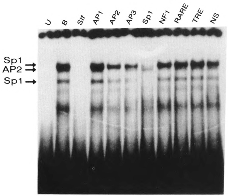
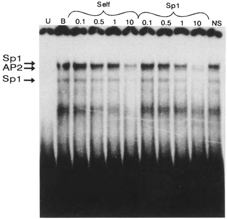
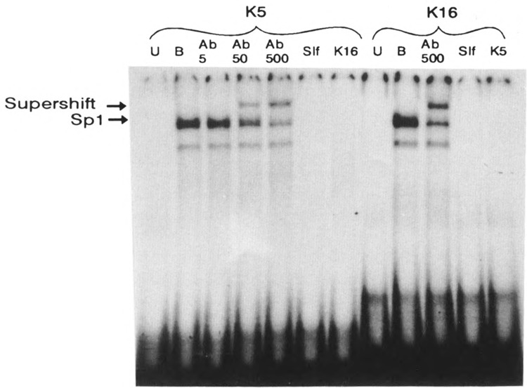
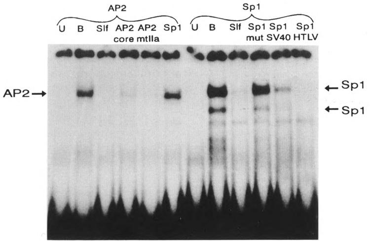
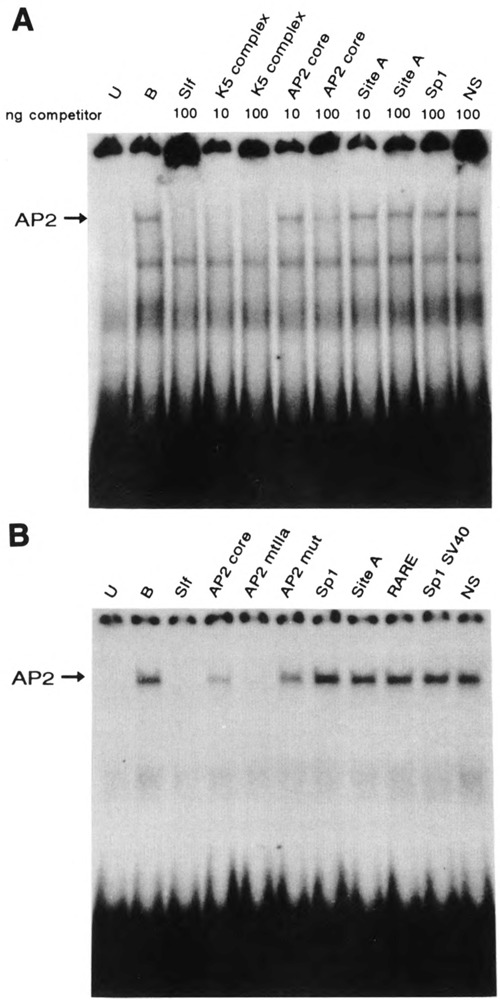
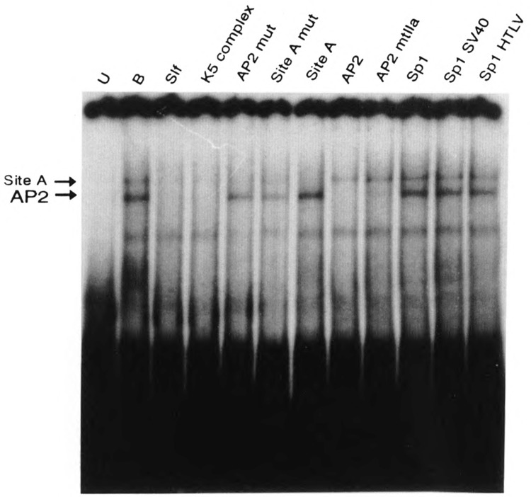
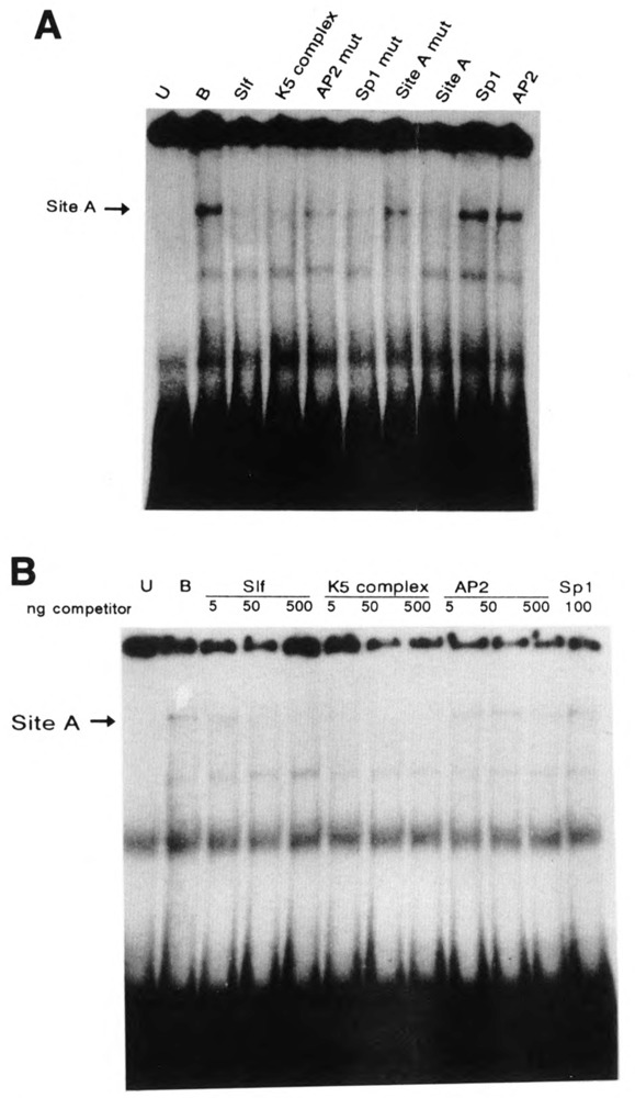
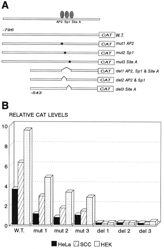
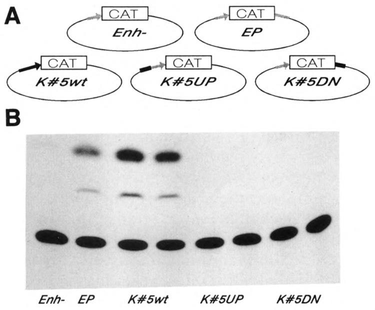
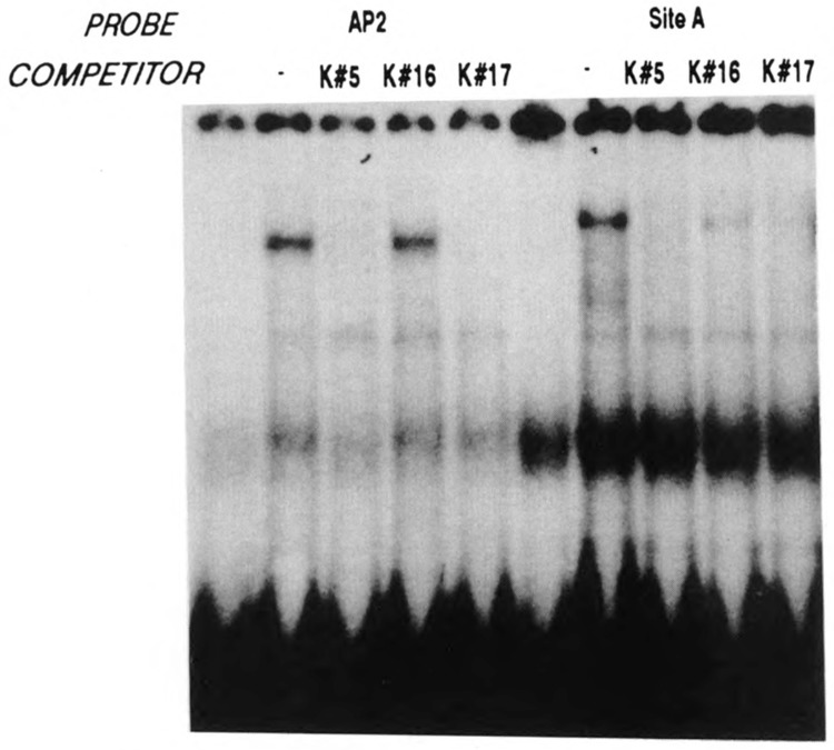
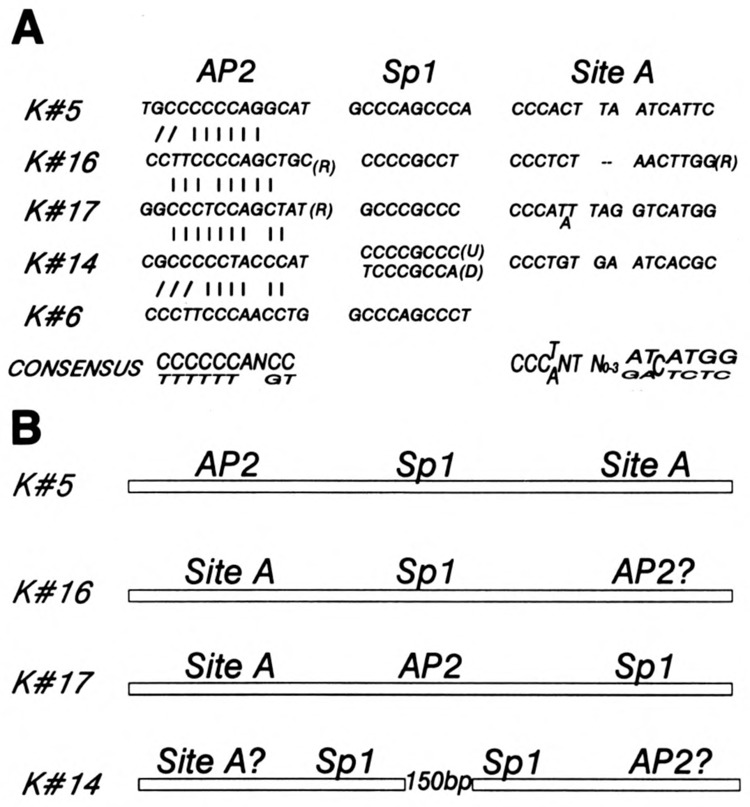
A common feature of all epithelial cells is the presence of keratin proteins that assemble into an intermediate filament cytoskeletal network. Whereas other cell types often use a specific master transcription factor to coordinate cell type-specific transcription, analysis of transcriptional regulation of keratin genes suggests that specific groupings of widely expressed transcription factors, acting on clusters of recognition elements in the promoter regions, confer epithelia-specific transcription. We define such a cluster of three sites that binds five transcription factors in the human K5 keratin gene. Within this cluster, an unusual Sp1 site binds the Sp1 transcription factor and two additional proteins. Flanking the Sp1 site are an AP2 site and another sequence, Site A; each binds a transcription factor. Similar clusters of recognition sites for the same five transcription factors have been also identified in other keratin genes. Such clusters may play a role in epithelia-specific expression of keratins.


Figures

Figure 1
Figure 1
The oligonucleotide probe that contains the K5 complex binds a large number of nuclear proteins. The first two lanes, U and B, contain the DNA probe unbound and bound to nuclear proteins, respectively, without any competitor. Competitors (100 ng each) include the probe itself (Slf), commercially available oligonucleotides containing recognition elements for various transcription factors, and synthetic recognition elements for the retinoic acid receptor (RARE) and the thyroid hormone receptor (TRE). NS, the nonspecific competitor, contains K5 promoter site B, located further upstream from the complex site (Ohtsuki et al., 1992).
Figure 2
Figure 2
The consensus Sp1 binding sequence is approximately as effective as the probe itself in competition for the binding of the transcription factor. This means that the protein binds to the unusual K5 gene sequence as well as to the consensus Sp1 sequence. The amounts of competitors range from 0.1 to 10 ng; 100 ng of the same nonspecific DNA competitor as in Figure 1 were added to lane NS.
Figure 3
Figure 3
Supershift experiment using Sp1-specific antibodies. The probes contained the Sp1 site from the K5 (left) or K16 gene (right). The lanes marked Ab contain the antibody, 5 to 500 ng, as indicated. Beside self-competition (Slf), each of the two probes competes with the other one as well (lanes marked K16 and K5).
Figure 4
Figure 4
The same three proteins that bind the Sp1 site are found both in HeLa cells and in primary human epidermal keratinocytes (HEK). Note that similar levels, both relative and absolute, of the bands are found in both nuclear extracts.
Figure 5
Figure 5
AP2 and Sp1 sites bind protein independently. The binding to the AP2 site, left, is abolished by the probe itself (Slf), i.e., the AP2 site of the K5 gene; by the core element of the AP2 site (core); and by the human metallothionein IIa gene promoter AP2 site (mtlla); but not by the Sp1 consensus sequence. Conversely, binding to the Sp1 site, right, is completed by the probe (Slf), i.e., the Sp1 site of the K5 gene, and also by the Sp1 sites from the SV40 and HTLV-1 promoters, but not by the mutated Sp1 site. Lanes U and B contain unbound and protein-bound probes without competitor DNA.
Figure 6
Figure 6
Characterization of the AP2 site. A. The competitors for binding include the probe itself (Slf), as well as the three-site complex and—less efficiently—the K5 AP2 core element. Neither Site A- nor Sp1-containing sequences competed for binding. B. The Mtlla gene AP2 site is as effective a competitor as the probe itself (Slf), i.e., the K5 gene AP2 site, whereas a mutated AP2 site, Sp1, Site A, RARE, and nonspecific DNA are not.
Figure 7
Figure 7
Binding to Sp1 obscures another element, Site A. However, with a probe containing a mutated Sp1 site, binding to Site A becomes clear. Both the mutant (Slf) and the wild-type K5 complex DNAs compete for binding. Various competitors specifically compete for binding either to Site A or to the AP2 site, whereas Sp1 site-containing oligonucleotides do not.
Figure 8
Figure 8
Binding to Site A is independent of the AP2 site. A. The probe contains point mutations both in the Sp1 and in the AP2 sites, leaving Site A as the only specific protein binding site. Note that only competitors containing an intact Site A compete for binding, whereas all other oligonucleotides fail to do so. B. The probe containing Site A and additional sequences downstream binds the same nuclear protein. Increasing amounts of the K5 complex compete for binding, but neither the AP2 nor the Sp1 site does. Oligonucleotides used as probes and competitors are listed in Tables 1 and 2.
Figure 9
Figure 9
Each of the three sites is important for the promoter function. A. Structures of the DNA constructs. Asterisks indicate point mutations. B. The constructs were transfected into three cell types, HeLa, squamous cell carcinoma line SCC-F12, and primary cultures of human epidermal keratinocytes. Each of the point mutations reduces the transcription levels two- to threefold in all three cell types. Deletions reduce CAT levels at least tenfold, to virtually unmeasurable.
Figure 10
Figure 10
The three-site complex is not an enhancer. A. Structures of the transfected DNAs, including the control construct containing both the enhancer and the promoter of the SV40 virus (EP), the construct containing 796 bp of the K5 promoter (K5WT), the enhancer trap vector (Enh-), and the two constructs containing the three-site complex either downstream from the CAT gene inserted into the BamH I site (K5DN), or upstream into the Bgl II site (K5UP). B. Results of the CAT assays. Duplicate transfections of constructs containing the K5 gene DNA are shown. Note that neither the K5UP nor the K5DN construct produces CAT activity higher than the parent enhancer trap vector.
Figure 11
Figure 11
K16 and K17 DNA both compete for binding at Site A, but only K17 at the AP2 site. The probe used on the left is the same as in Figure 6B; the one on the right is the longer Site A probe presented in Figure 8B. In both cases, the first lane contains no nuclear protein.
Figure 12
Figure 12
Common protein binding sites in promoters of keratin genes. A. Sequences of the sites. R marks sequences found in the reverse orientation. Vertical lines in the AP2 segments indicate identical nucleotides; slanted lines mark the additional runs of C residues. Note two different Sp1 sequences. U and D mark the upstream and the downstream consensus Sp1 sites in the K14 gene promoter. Allowing various numbers of bases within Site A gives improved alignment of sequences. B. Arrangement of sites within the promoters. The tightest arrangement is in the K5 gene. In the K14 gene, the two areas are separated by approximately 150 bp. Question marks indicate that these sites have been identified by sequence comparison only, and not yet by gel shift or mutagenesis experiments.

Similar articles

Cited by

References

    1. Anderson G. M. and Freytag S. O. (1991), Mol Cell Biol 11, 1935–1943. - PMC - PubMed
    1. Bernerd F., Magnaldo T., Freedberg I. M., and Blumenberg M. (1993), Gene Expr 3, 187–199. - PMC - PubMed
    1. Blessing M., Zentgraf H., and Jorcano J. L. (1987), EMBO J 6, 567–575. - PMC - PubMed
    1. Briggs M. R., Kadonaga J. T., Bell S. P., and Tjian R. (1986), Science 234, 47–52. - PubMed
    1. Chen C. and Okayama H. (1987), Mol Cell Biol 7, 2745–2752. - PMC - PubMed

Publication types