. 1994 Feb 15;8(4):440-52.
doi: 10.1101/gad.8.4.440.

Distinct molecular mechanism regulate cell cycle timing at successive stages of Drosophila embryogenesis

Affiliations

Distinct molecular mechanism regulate cell cycle timing at successive stages of Drosophila embryogenesis

B A Edgar et al. Genes Dev. .

Abstract

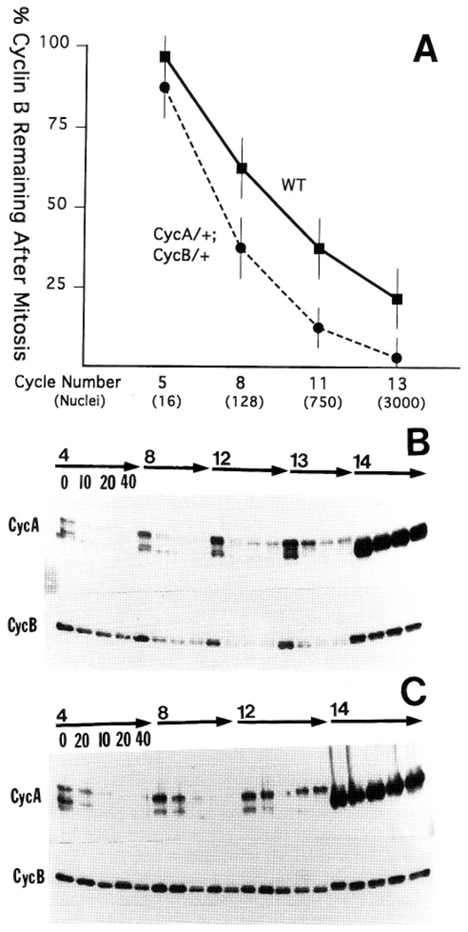
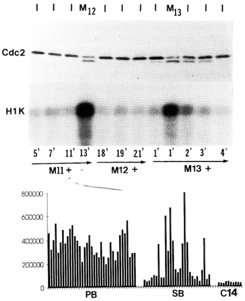
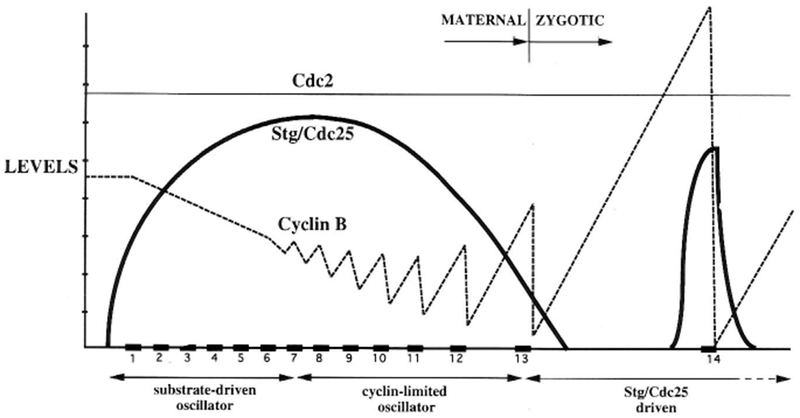
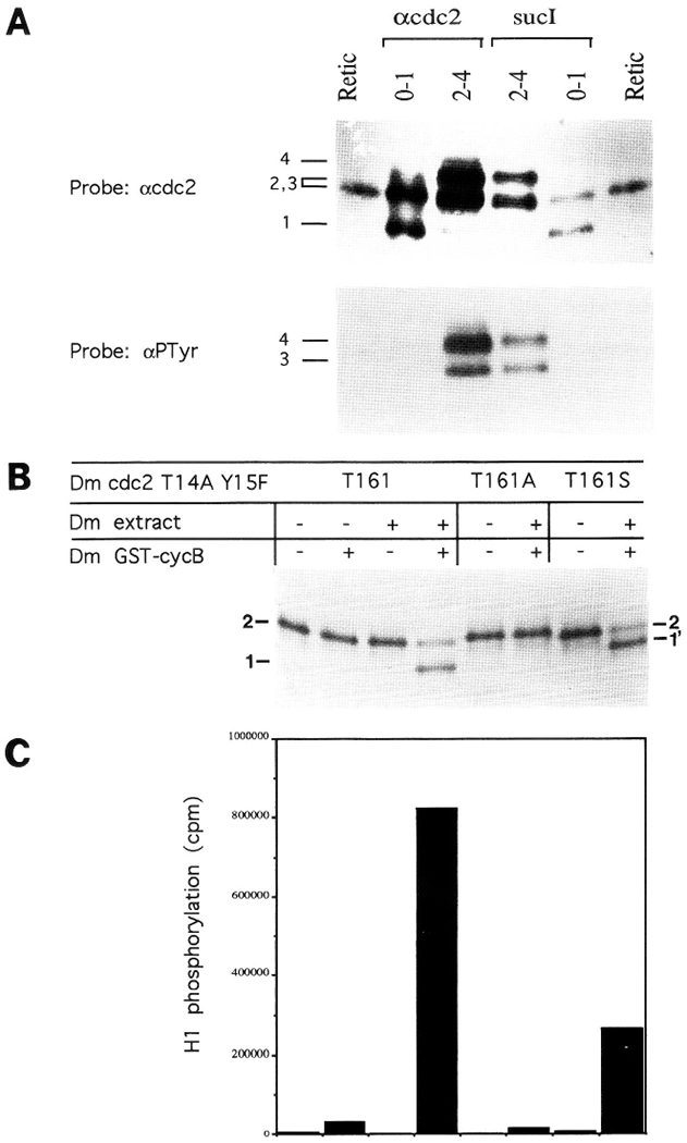
The conserved regulators of cell cycle progression--Cyclins, Cdc2 kinase, and String phosphatase (Cdc25)--accommodate multiple modes of regulation during Drosophila embryogenesis. During cell cycles 2-7, Cdc2/Cyclin complexes are continuously present and show little fluctuation in abundance, phosphomodification, or activity. This suggests that cycling of the mitotic apparatus does not require cytoplasmic oscillations of known regulatory activities. During cycles 8-13 a progressive increase in the degradation of Cyclins at mitosis leads to increasing oscillations of Cdc2 kinase activity. Mutants deficient in cyclin mRNAs suffer cell cycle delays during this period, suggesting that Cyclin accumulation times these cycles. During interphase 14, programmed degradation of maternal String protein leads to inhibitory phosphorylation of Cdc2 and cell cycle arrest. Subsequently, mitoses 14-16 are triggered by pulses of zygotic string transcription.


Figures

Figure 1.
Figure 1.
Spatial expression of String protein during embryogenesis. (AE) Embryos at progressively more advanced stages of development, immunohistochemically stained for String protein, and photographed using DIC optics. (A) Nuclear cycle 11: Note the concentration of String in nuclei. High magnification observations revealed that String is concentrated in nuclei during the syncytial interphases and on chromosomes during all stages of mitosis (not shown). This embryo was stained severalfold less intensely than the others to highlight this nuclear staining. (B) Interphase 14, maternal String protein is completely degraded. (C) Early mitosis 14, zygotically expressed String appears in mitotic domains 1–11 (Foe 1989). (D) Patterns of mitosis 15. (E) String expression in proliferating neuroblasts of the central and peripheral nervous systems during postcycle 16 embryogenesis.
Figure 2.
Figure 2.
Regulation of Cdc2 by phosphorylation during cycles 13 and 14. (A) Time-course immunoblot of Cdc2 from metaphase and anaphase of cycle 13 (M13, A13) through S phase, G2, and mitosis of cycle 14. Methanol-fixed embryos were staged as described in Fig. 3 and Materials and methods. S phase and G2 time points were ordered by the degree of cellularization (McKnight and Miller 1977; Edgar and Schubiger 1986.) The four phosphoisoforms of Cdc2 can be distinguished in the M14 lane, and are labeled (right, 1–4). Active T161-phospho-Cdc2 (form 1) is the fastest migrating, unphosphorylated Cdc2 is above this (form 2), and the two tyrosine-phosphorylated forms (Y15- and Y15, T14-phospho) are slowest migrating (forms 3 and 4; Fig. 8). Active Cdc2 disappears during early S phase 14, and is replaced by tyrosine-phosphorylated forms. During mitosis 14, active T161-phospho-Cdc2 reappears, and during the first G1 period (G117), Cdc2 is unphosphorylated. (B) Immunoblots of wild-type embryos in G214 (WT), similarly aged embryos (genotype stg7B hsstg/stg7B hs–stg), which were driven into mitosis 14 by expression of String from the heat shock promotor (hs–stg), and embryos of the genotype stg7B/stg7B arrested in G214 by the string mutation (stg). Note that induction of String causes tyrosine dephosphorylation Cdc2, leading to increased mobility. (BG) Background bands.
Figure 3.
Figure 3.
Mitotic synchrony and staging of methanol-fixed embryos. Each panel shows a field that covers ~80% of the surface of a methanol-fixed, Hoecht 33258-stained cycle 10 embryo, such as those used in our immunoblotting experiments. Only the poles of the embryos (which would be out of the focal plane) are not shown; however, nuclei in the poles exhibit the same synchrony as those shown. Between early interphase (EI) and late interphase (LI), nuclei increase their volume several fold. At prophase (P), chromatin condenses onto the nuclear envelope; at metaphase (M), condensed chromosomes congregate to form the metaphase plate. Anaphases (A) and telophases (T) also occur synchronously.
Figure 4.
Figure 4.
Changes in cell cycle regulators during cycles 2–7 (A) and 8–14 (B). We show time-course immunoblots probed with antibodies to String (Stg|, Cyclin A (CycA), Cyclin B (CycB), and Cdc2 (Cdc2). Cycle numbers and phases are denoted (top). The embryos were precisely staged as interphase (I), prophase (P), metaphase (M), anaphase (A) or telophase (T). In later cycles, several interphase time points were ordered using nuclear volume, which increases with time (Fig. 3). Phosphatase treatment indicated that mobility shifts of String are due to relative hyperphosphorylation during mitosis. Lower mobility bands of Cyclin A are likewise due to phosphorylation (see Materials and methods). Phosphorylation of Cdc2 at T161 causes a downshift, whereas Y15 and T14 phosphorylation cause upshifts (in cycle 14 only; see Materials and methods and Fig. 8). Both unphosphorylated and T161-phospho-Cdc2 are continuously present during cycles 2–9. One embryo was loaded per lane in A, and three embryos of identical stage were loaded in B. The same blots were serially probed with each antibody, except for String in A, which was from a separate experiment. These experiments were repeated four times (A) or six times (B), with similar results.
Figure 5.
Figure 5.
(A) Mitotic degradation of Cyclin B increases as the nuclei proliferate. Cyclin B levels were measured by scanning densitometry of dilution-series immunoblots. Metaphase and anaphase levels for each of cycles 5, 8, 11, and 13 were measured, using samples containing 10 embryos of identical stage. (WT) Wild-type embryos; (CycA/+; CycB/+ ) embryos from mothers heterozygous for a cyclin A mutation (neo114) and a deficiency covering the cyclin B gene (Df(2R)59AD). These embryos receive half the normal dose of cyclin mRNA. Standard deviations were derived from multiple loadings and exposures of a sample, and levels were normalized to Cdc2, a stable protein. (B) Cyclin turnover in embryos arrested in interphase by cycloheximide. Permeabilized embryos were treated with 20 μg/ml of cycloheximide for 0, 10, 20, and 40 min and then methanol-fixed. Three embryos arrested in interphase of each of cycles 4, 8, 12, 13, and 14 were selected, pooled, and subjected to immunoblot analysis. Numbers above the arrows indicate the cycle of arrest, and numbers below the arrows indicate minutes of treatment with cycloheximide (these follow the same order for each cycle displayed). Note that Cyclin A (CycA) is degraded rapidly and completely after arrest during the earlier cycles and that degradation becomes less complete in later cycles. In contrast, a greater proportion of Cyclin B (CycB) is degraded with progressively later cycles. Both Cyclins are stable during interphase 14. The 0 min time points represent late interphase embryos that were not treated with cycloheximide. (C) Cyclin stability in embryos arrested in metaphase by colcemid. Permeabilized embryos were first treated with 20 μg/ml of colcemid for 20 min. This depolymerizes microtubules and results in metaphase arrest. Following the 20-min colcemid treatment, embryos were treated with 20 μg/ml of cycloheximide and 20 μg/ml of colcemid for 10, 20, or 40 min to assess the stability of the Cyclins during metaphase arrest. The embryos were then methanol-fixed, and three embryos arrested in metaphases 4, 8, 12, and interphase 14 from each time point were selected, pooled, and subjected to immunoblot analysis. Note that Cyclin A is unstable during metaphase arrest, whereas Cyclin B is essentially stable. 0-Min time points represent untreated metaphase embryos (except for the cycle 14 time points, which are all interphase embryos).
Figure 6.
Figure 6.
Cdc2 kinase activity oscillates in synchrony with T161-phospho-Cdc2 during the syncytial blastoderm cycles, but does not oscillate during the preblastoderm cycles. (Top) Both the state of Cdc2 and its kinase activity in single embryos are shown from cycles 12, 13, and early 14. We staged individual living embryos visually, keeping track of the minutes elapsed between mitosis and the time of lysis (noted below lower gel). Mitoses were scored by the disappearance of the nuclear envelope using DIC optics, and cycle numbers were determined using nuclear density (Foe and Alberts 1983). The cycle phase of each embryo at the time of lysis is noted along the top (I = interphase, M = mitosis). Half of each embryo lysate was immunoblotted for Cdc2 (upper gel; Cdc2), and the other half was tested for kinase activity after immunoprecipitation with anti-Cdc2 antisera (lower; H1K). Note that high levels of Cdc2-associated H1 kinase activity are confined to those embryos with the faster-migrating form of Cdc2, which is T161 phosphorylated (Fig. 8). This experiment was repeated four times with similar results. (Below) We show the H1 kinase activity of Cdc2 immunoprecipitates of 38 random single preblastoderm embryos (PB; cycles 2–8), 28 random syncytial blastoderm embryos (SB; cycles 10–13), and 10 interphase 14 (C14) embryos. H1 kinase activity was quantified by Phosphorimaging, after SDS–gel electrophoresis and electroblotting. Note that Cdc2 kinase activity fluctuates little in the preblastoderm embryos, fluctuates dramatically in the syncytial blastoderm embryos, and is uniformly low in interphase 14 embryos.
Figure 7.
Figure 7.
Summary, showing fluctuations in levels of String (bold line), Cyclin B (dashed line), and Cdc2 (fine line) during the first 14 cell cycles. Protein levels are depicted along the y-axis on a linear scale, with arbitrary units. Time is indicated along the x-axis, with periods of mitosis boxed in black and numbered. During cycles 1–7, there is little fluctuation in Cyclin levels or Cdc2 kinase activity. Beginning at cycle 8, Cyclin degradation at metaphase/anaphase transitions becomes apparent, and the cycles begin to slow. At this point Cdc2 kinase activity also begins to fluctuate, due to gain and loss of the activating phosphate at T161. It is note-worthy that greater amounts of Cyclin are degraded with progressively later cycles. Because nuclei and mitotic apparatuses (presumed Cdc2/Cyclin substrates) increase exponentially in number, this suggests a connection between Cyclin utilization and its degradation. Accordingly, we propose that cycle slowdown following mitosis 8 is effected by titration and depletion of maternal Cyclins (Table 1). Mitosis 14 is timed by a distinct mechanism, in which inactive Cdc2/Cyclin complexes are activated by String translated from new zygotic transcripts.
Figure 8.
Figure 8.
Identification of Cdc2 phosphoisoforms. (A) Tyrosine phosphorylation of Cdc2 results in reduced electrophoretic mobility. Cdc2 was precipitated from 0- to 1-hr (cycles 1–6) or 2- to 4-hr (cycles 13–15) embryos using either anti-Cdc2 antisera or sucl beads (top). The precipitated proteins, along with in vitro-translated Cdc2 (Retie) were immunoblotted with either anti-Cdc2 antibodies (top) or anti-phosphotyrosine antibodies (lower). Note that the two upper bands (forms 3 and 4) in the 2- to 4-hr samples contain phosphotyrosine, and that no phosphotyrosine is found in the two faster migrating Cdc2 isoforms (forms 1 and 2) found in 0- to 1-hr embryos. In this gel, forms 2 and 3 comigrated (see Fig. 2A for separation). Numbers referring to the different Cdc2 forms (1–4) are to the left of the gels. (B) Phosphorylation at T161 increases Cdc2 mobility. HA-tagged, 35S-labeled, in vitro-translated Drosophila Cdc2 containing nonphosphory-latable amino acid substitutions at T14 (A) and Y15 (F) was incubated in the presence (+) or absence (−) of extracts of early Drosophila embryos, and GST–CyclinB (above). The labeled protein was then analyzed by SDS-PAGE. Both Cyclin B and the embryo extract, which contains a T161 kinase activity, are required to generate the downshifted isoform (form 1). When T161 is mutated to the non-phosphorylatable residue alanine (T161A), this downshift does not occur; but when T161 is replaced by phosphorylatable serine (T161S) a somewhat less extreme downshift occurs (1′). (C) Phospho-T161-Cdc2 (form 1) is the active H1 kinase. A fraction of each of the samples shown in B was immunoprecipitated using anti-HA antibodies, and its kinase activity was measured using histone H1 as a substrate. The order of samples in B and C is the same. Only samples containing the down-shifted (form 1) Cdc2 have high kinase activity. See Fig. 6 for the analogous experiment in vivo.

References

    1. Alfa CE, Ducommun B, Beach D, and Hyams JS. 1990. Distinct nuclear and spindle pole body population of cyclin-cdc2 in fission yeast. Nature 347: 680–682. - PubMed
    1. Amon A, Surana U, Muroff I, and Nasmyth K. 1992. Regulation of p34CDC28 tyrosine phosphorylation is not required for entry into mitosis in S. cerevisiae. Nature 355: 368–371. - PubMed
    1. Booher R and Beach D. 1988. Involvement of cdc13+ in mitotic control in Schizosaccharomyces pombe; Possible interaction of the gene product with microtubules. EMBO J. 6: 3441–3447. - PMC - PubMed
    1. Clarke PR, Leiss D, Pagano M, and Karsenti E. 1992. Cyclin A- and Cyclin B-dependent kinases are regulated by different mechanisms in Xenopus egg extracts. EMBO J. 11: 1751–1761. - PMC - PubMed
    1. Davis FM, Tsao TY, Fowler SK, and Roa PN. 1983. Monoclonal antibodies to mitotic cells. Proc. Nat. Acad. Sci 80: 2926–2930. - PMC - PubMed

Publication types