. 1994 Sep;120(9):2687-702.
doi: 10.1242/dev.120.9.2687.

Specific roles of the alpha V beta 1, alpha V beta 3 and alpha V beta 5 integrins in avian neural crest cell adhesion and migration on vitronectin

Affiliations

Specific roles of the alpha V beta 1, alpha V beta 3 and alpha V beta 5 integrins in avian neural crest cell adhesion and migration on vitronectin

M Delannet et al. Development. 1994 Sep.

Abstract

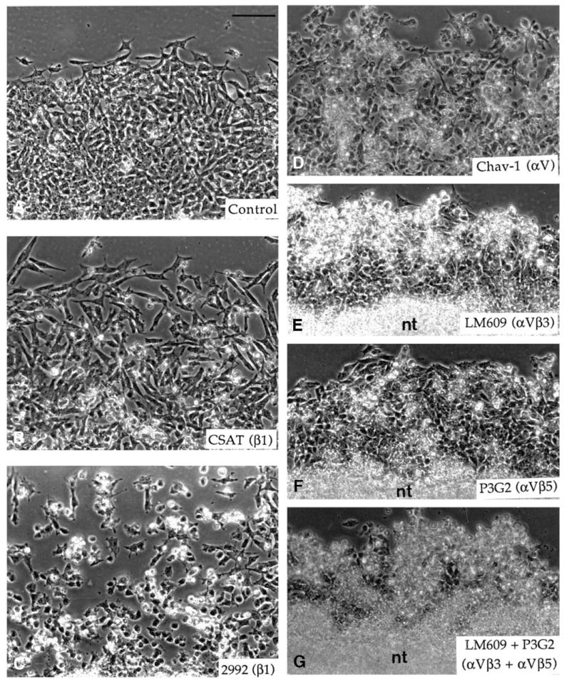
To identify potentially important extracellular matrix adhesive molecules in neural crest cell migration, the possible role of vitronectin and its corresponding integrin receptors was examined in the adhesion and migration of avian neural crest cells in vitro. Adhesion and migration on vitronectin were comparable to those found on fibronectin and could be almost entirely abolished by antibodies against vitronectin and by RGD peptides. Immunoprecipitation and immunocytochemistry analyses revealed that neural crest cells expressed primarily the alpha V beta 1, alpha V beta 3 and alpha V beta 5 integrins as possible vitronectin receptors. Inhibition assays of cellular adhesion and migration with function-perturbing antibodies demonstrated that adhesion of neural crest cells to vitronectin was mediated essentially by one or more of the different alpha V integrins, with a possible preeminence of alpha V beta 1, whereas cell migration involved mostly the alpha V beta 3 and alpha V beta 5 integrins. Immunofluorescence labeling of cultured motile neural crest cells revealed that the alpha V integrins are differentially distributed on the cell surface. The beta 1 and alpha V subunits were both diffuse on the surface of cells and in focal adhesion sites in association with vinculin, talin and alpha-actinin, whereas the alpha V beta 3 and alpha V beta 5 integrins were essentially diffuse on the cell surface. Finally, vitronectin could be detected by immunoblotting and immunohistochemistry in the early embryo during the ontogeny of the neural crest. It was in particular closely associated with the surface of migrating neural crest cells. In conclusion, our study indicates that neural crest cells can adhere to and migrate on vitronectin in vitro by an RGD-dependent mechanism involving at least the alpha V beta 1, alpha V beta 3 and alpha V beta 5 integrins and that these integrins may have specific roles in the control of cell adhesion and migration.


Figures

Fig. 1
Fig. 1
Immunoblotting analysis of vitronectin expression in embryos at the time of neural crest cell development. Extracts of truncal and cranial regions of embryos at E2.5 (lanes 1 and 2, respectively) and of truncal regions of embryos at E3.5 and E4.5 (lanes 3 and 4, respectively) were subjected to immunoblotting using a monoclonal antibody to human vitronectin. Approximately the same amounts of material were loaded in the different lanes. Two major bands of about Mr of 68K and 43K are found at very similar amounts in all embryo extracts. The upper band corresponds to vitronectin and the lower band corresponds either to a degradation product of vitronectin or to one of the forms of yolk vitronectin (Nagano et al., 1992). The band of Mr of 78K that can be seen in lane 4 is likely can be attributed to the high molecular mass form of vitronectin (Kitagaki-Ogawa et al., 1990). Molecular mass standards are indicated on the left.
Fig. 2
Fig. 2
Immunofluorescence analysis of the distribution of vitronectin at the time of neural crest cell migration. (A,B) Consecutive transverse sections through the brachial level of a 25-somite-stage embryo (E2.5) stained for vitronectin and fibronectin, respectively. Vitronectin immunoreactivity is present on the surface of all cells in the embryo. Staining is also prominent on neural crest cells (indicated by arrows). (C,D) Consecutive transverse sections through the rostral part of a sclerotome at the brachial level of a 32-somite stage embryo (E3-3.5) stained for vitronectin and NC-1, respectively. Both neural crest cells accumulating along the neural tube to form the spinal ganglion and those migrating through the sclerotome (indicated by arrows) show a strong reactivity for vitronectin. a, aorta; d, dermamyotome; e, ectoderm; n, notochord; nc, neural crest cells; nt, neural tube; s, somite; sc, sclerotome. (A,B, ×230; C,D, ×220).
Fig. 3
Fig. 3
Immunofluorescence analysis of the distribution of vitronectin at the time of formation of peripheral ganglia. (A,B) Transverse sections through the dorsal root ganglion at the brachial level of embryos at E5 and E9 stained for vitronectin. The spinal cord, motor nerves and neurons of the dorsal root ganglion show a strong immunoreactivity for vitronectin at E5, but are faintly labeled at E9, i.e. after neuronal differentiation has occurred. g, dorsal root ganglion; n, motor nerve; s, spinal cord. (A, ×180; B, ×160).
Fig. 4
Fig. 4
Comparative attachment (A), spreading (B) and migration (C) of neural crest cells on vitronectin (closed squares) and fibronectin (open circles) adsorbed onto bacteriological Petri dishes at concentrations ranging from 0.05 μg/ml to 100 μg/ml. Migration assays were performed in serum-free medium. In A and B, results are expressed as the percentage of attached and spread cells in relation to the total number of cells deposited on the substratum. In C, results are expressed as the linear distance in μm between the periphery of the neural crest outgrowth and the edge of the neural tube explant after 18 hours of culture. Each value represents, in A and B, the mean ± s.d. of at least 6 different measurements in at least 3 independent experiments and in C the mean ± s.d. of at least 20 different measurements in at least 3 independent experiments.
Fig. 5
Fig. 5
Migration of neural crest cells on vitronectin adsorbed to the substratum at the concentrations of 50 μg/ml (A,B) and 0.5 μg/ml (C,D). (B,D) Higher magnifications of A and C, respectively, showing the morphology of cells. Neural tube explants were deposited onto vitronectin-coated bacteriological Petri dishes and neural crest cells were allowed to migrate on the substratum for 15 hours in serum-free medium and then photographed. At high coating concentrations, vitronectin yields a large number of well-spread cells that migrate long distances from the neural tube. At low concentrations, vitronectin also produces significant migration of cells, though most of them are poorly spread. nt, neural tube.
Fig. 6
Fig. 6
Neural crest cell attachment (A), spreading (B) and migration (C) on vitronectin at 5-10 μg/ml in the presence of antibodies to vitronectin at the concentrations of 1 mg/ml and 0.1 mg/ml or to fibronectin at 1 mg/ml, and of RGDS peptides at the concentrations of 1 mg/ml and 0.1 mg/ml. In A and B, results are expressed as the percentage of attachment and spreading in control experiments. In control assays, at least 50% of the cells deposited on the substratum were adherent. In C, neural crest cells were allowed to emigrate from the neural tube explants for 5 hours, and antibodies or peptides were then applied to the culture for 15 hours in DMEM supplemented with 5% serum. Results are expressed as the linear distance in μm between the periphery of the neural crest outgrowth and the edge of the neural tube explant after 15 hours of culture in the presence of the antibodies or peptides. Each value represents in A and B the mean ± s.d. of at least six different measurements in at least three independent experiments and in C the mean ± s.d. of at least 20 different measurements in at least 3 independent experiments.
Fig. 7
Fig. 7
Migration of neural crest cells on vitronectin in the presence of antibodies to vitronectin at the concentration of 1 mg/ml (A), of antibodies to fibronectin at 1 mg/ml (B) and of RGDS peptides at 1 mg/ml (C). Neural crest cells were allowed to emigrate from the neural tube explants on vitronectin at 10 μg/ml for 5 hours. Antibodies or peptides were then applied to the culture for 4 hours. Both the antibodies to vitronectin and the RGDS peptides produce extensive rounding up of neural crest cells followed by cell aggregation. nt, neural tube. Bar, 100 μm.
Fig. 8
Fig. 8
(A) Vitronectin synthesis by neural crest cells. Cell extract (lane 1) and culture supernatant (lane 2) of metabolically labeled neural crest cells were subjected to immunoprecipitation with antibodies to vitronectin. (B) Integrin receptors for vitronectin on neural crest cells. Neural crest cells cultured on vitronectin were metabolically labeled, extracted in detergent and the lysates were subjected to immunoprecipitation using the VNR-C3 polyclonal antibody to the cytoplasmic domain of the chicken αV integrin subunit (lane 1), the 2992 polyclonal antibody to the chicken β1 integrin subunit (lane 2), the LM609 monoclonal antibody to the human αVβ3 integrin complex (lane 3) and the 4377 polyclonal antibody to the cytoplasmic domain of the human β5 integrin subunit (lane 4). Samples were resolved by SDS-PAGE on 10% acrylamide gels under reducing conditions in A and on 7.5% acrylamide gels under nonreducing conditions in B and radiolabeled bands were visualized by fluorography. Molecular mass markers are indicated on the left of panels A and B.
Fig. 9
Fig. 9
Neural crest cell spreading (A) and migration (B) on vitronectin adsorbed onto bacteriological Petri dishes at 5 μg/ml in the presence of various function-perturbing antibodies to integrins. The polyclonal antibody 2992 (anti-β1), mAb CSAT (anti-β1), mAb Chav-1 (anti-αV), mAb LM609 (anti-αVβ3) and mAb P3G2 (anti-αVβ5) were used in adhesion assays at 1 mg/ml, 100 μg/ml, 25 μg/ml, 10 μg/ml and 10 μg/ml, respectively. In migration assays, the polyclonal antibody 2992 was at 1 mg/ml and the various mAbs were at 50 μg/ml. Results are expressed as the percentage of spreading and migration in control experiments. In control assays for cellular adhesion, at least 50% of the cells deposited on the substratum were adherent. In assays for cellular migration, neural crest cells were allowed to emigrate from the neural tube explants for 5 hours and antibodies were then added to the culture medium for 15 hours. Each value represents, in A, the mean ± s.d. of at least 6 different measurements in at least 3 independent experiments and, in B, the mean ± s.d. of at least 20 different measurements in at least 3 independent experiments.
Fig. 10
Fig. 10
Migration of neural crest cells on vitronectin in the presence of various function-perturbing antibodies to integrins. Neural crest cells were allowed to emigrate from the neural tube explants on vitronectin at the concentration of 10 μg/ml for 5 hours. Antibodies were then applied to the culture for 15 hours. (A) Control without antibody, (B) mAb CSAT (anti-β1) at 250 μg/ml, (C) polyclonal antibody 2992 (anti-β1) at 0.5 mg/ml, (D) mAb Chav-1 (anti-αV) at 50 μg/ml, (E) mAb LM609 (anti-αVβ3) at 50 μg/ml, (F) mAb P3G2 (anti-αVβ5) at 50 μg/ml and (G) mAbs LM609 and P3G2 both at 50 μg/ml. nt, neural tube. Bar, 100 μm.
Fig. 11
Fig. 11
Immunofluorescence detection of the β1 (A) and αV (B) integrin subunits, the αVβ3 (C) and αVβ5 (D) integrins, vinculin (E) and α-actinin (F) on neural crest cells cultured on vitronectin. Cells were cultured for 18 hours and fixed using different procedures before immunofluorescence staining. For the β1 subunit, cells were fixed in PBS containing 3.7% formaldehyde, 0.5% Triton X-100 and 5% sucrose for 5 minutes and postfixed in 3.7% formaldehyde for 1 hour. For the αV subunit, cells were fixed in cold methanol for 5 minutes followed by cold acetone for 1 minute. For vinculin and α-actinin, cells were first fixed in 3.7% formaldehyde for 1 hour and permeabilized with 0.5% Triton X-100 during 5 minutes. For the αVβ3 and αVβ5 integrins, cells were first fixed in 3.7% formaldehyde for 1 hour. Neural crest cells exhibit β1 and αV integrin subunits both in focal adhesion sites and as a diffuse pattern on their surface. The αVβ3 and αVβ5 integrin receptors display essentially a uniform, diffuse pattern of staining on the cell surface and are not concentrated in adhesion plaques. Vinculin is concentrated in focal contacts at the tips of cell processes and α-actinin is also in focal contacts and along actin microfilaments. Bar, 25 μm.

References

    1. Akiyama SK, Yamada SS, Yamada KM. Characterization of a 140-kD avian cell surface antigen as a fibronectin-binding molecule. J Cell Biol. 1986;102:442–448. - PMC - PubMed
    1. Barnes DW, Reing JE, Amos B. Heparin-binding properties of human serum spreading factor. J Biol Chem. 1985;260:9117–9122. - PubMed
    1. Bodary SC, McLean JW. The integrin β1 subunit associates with the vitronectin receptor αv subunit to form a novel vitronectin receptor in a human embryonic kidney cell line. J Biol Chem. 1990;265:5938–5941. - PubMed
    1. Bossy B, Reichardt LF. Chick integrin αV subunit molecular analysis reveals high conservation of structural domains and association with multiple β subunits in embryo fibroblasts. Biochemistry. 1990;29:10191–10198. - PMC - PubMed
    1. Boucaut J-C, Darribère T, Poole TJ, Aoyama H, Yamada KM, Thiery JP. Biological active synthetic peptides as probes of embryonic development: A competitive peptide inhibitor of fibronectin function inhibits gastrulation in amphibian embryos and neural crest cell migration in avian embryos. J Cell Biol. 1984;99:1822–1830. - PMC - PubMed

Publication types