. 1995 Feb 10;267(5199):891-3.
doi: 10.1126/science.7531366.

Suppression of ICE and apoptosis in mammary epithelial cells by extracellular matrix

Affiliations

Suppression of ICE and apoptosis in mammary epithelial cells by extracellular matrix

N Boudreau et al. Science. .

Abstract

Apoptosis (programmed cell death) plays a major role in development and tissue regeneration. Basement membrane extracellular matrix (ECM), but not fibronectin or collagen, was shown to suppress apoptosis of mammary epithelial cells in tissue culture and in vivo. Apoptosis was induced by antibodies to beta 1 integrins or by overexpression of stromelysin-1, which degrades ECM. Expression of interleukin-1 beta converting enzyme (ICE) correlated with the loss of ECM, and inhibitors of ICE activity prevented apoptosis. These results suggest that ECM regulates apoptosis in mammary epithelial cells through an integrin-dependent negative regulation of ICE expression.


Figures
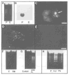
Fig. 1
Fig. 1
Characteristics of apoptosis in CID-9 cells. (A) Electrophoretic analysis of total DNA (20 μg) from cells cultured for 7 days on plastic (P) or EHS basement membrane (E). (B) RNA blot hybridized with a probe that detects the 2.4-kb mRNA for the apoptosis-associated gene SGP-2. (C) Acridine orange staining of cultured cells on plastic (arrow points to apoptotic cells). Scale bar, 58 μm. In situ analysis of DNA fragmentation in individual cells cultured on plastic (D) or EHS basement membrane (E) detected by fluorescein isothiocyanate–digoxigenin nucleotide labeling of 3′-OH DNA ends (Apoptag, Oncor). Scale bar, 90 μm. Electrophoretic analysis of total DNA (20 μg) from CID-9 cells (F) cultured on EHS (E) or allowed to form endogenous basement membrane (BM) for 5 days, (G) treated with normal rabbit serum (control) or anti–β1 integrin for 2 days, or (H) cultured on plastic (P), type I collagen (200 μg/ml) (Col), or fibronectin (50 μg/ml) (FN) for 5 days.
Fig. 2
Fig. 2
Apoptosis in cells overexpressing stromelysin-1. (A) Electrophoretic analysis of total DNA (20 μg) from control (C) and IPTG-induced cells (I) after 72 hours. Corresponding RNA blot (20 μg per lane) hybridization with a probe that detects a 1.9-kb stromelysin-1 mRNA. In situ analysis of DNA from mammary gland of normal mice in midpregnancy (B) and in transgenics expressing stromelysin-1 (14) (C). Note the increase in the number of epithelial cells undergoing apoptosis and the collapsed alveoli in the transgenics compared to normal mice. Scale bar, 33 μm.
Fig. 3
Fig. 3
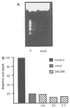
Inhibition of apoptosis in CID-9 cells. (A) Electrophoretic analysis of DNA (20 μg) from CID-9 untransfected control cells (P) or cells transfected with crmA and cultured on plastic for 7 days. (B) Quantitation of fragmented DNA from untransfected CID-9 cells (control), cells transfected with crmA, or cells treated with 0.5, 3.5, or 5.0 μM BACMK.
Fig. 4
Fig. 4
ICE mRNA and protein expression in mammary epithelium. (A) RNA blot (20 μg per lane) hybridized with a probe that detects a 1.6-kb ICE mRNA in mammary tissue from normal mice lactating for 9 days (L) or after involution for 2, 4, and 8 days (2l, 4l, 8l). (B) RNA blot for ICE mRNA in CID-9 cells after 5 days of culture on ECM (E) or tissue culture plastic (P). (C) Immunoblot analysis of ICE protein in lysates from corresponding cells with a polyclonal antibody that detects the 45-kD precursor, the active 20-kD subunit, and processing intermediates (19).

References

    1. Hentgartner MO, Horvitz HR. Cell. 1994;76:665. - PubMed
    2. Korsmeyer SJ. Cancer Surv. 1992;5:105. - PubMed
    1. Yuan J, et al. Cell. 1993;75:641. - PubMed
    2. Miura M, et al. ibid. :653.
    1. Gagliardini V, et al. Science. 1994;263:826. - PubMed
    1. Lin C, Bissell MJ. FASEB J. 1993;7:737. - PubMed
    2. Li ML, et al. Proc Natl Acad Sci USA. 1987;84:136. - PubMed
    1. Talhouk R, et al. J Cell Biol. 1992;118:1271. - PMC - PubMed
    2. Talhouk R, et al. Development. 1991;112:439. - PMC - PubMed

Publication types

MeSH terms