

Quantitative correlation between HLA class I allele expression and recognition of melanoma cells by antigen-specific cytotoxic T lymphocytes
- PMID: 7541714
- PMCID: PMC2248458
Quantitative correlation between HLA class I allele expression and recognition of melanoma cells by antigen-specific cytotoxic T lymphocytes
Abstract
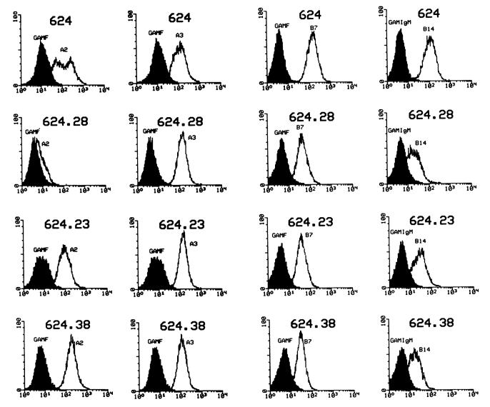
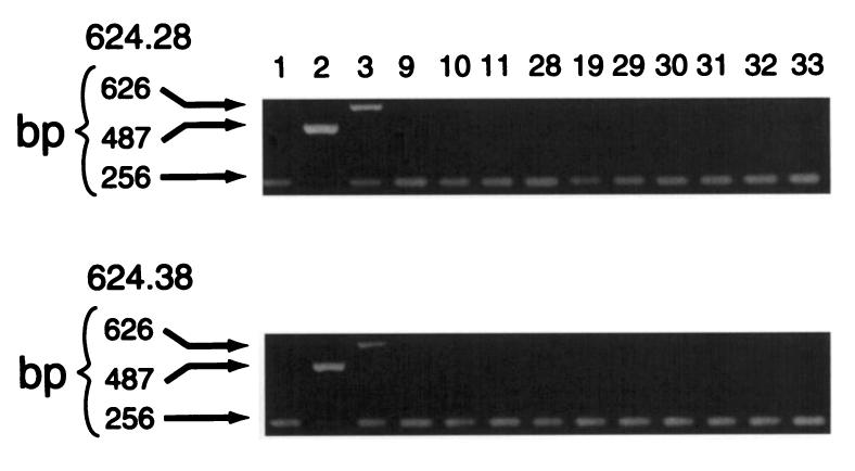
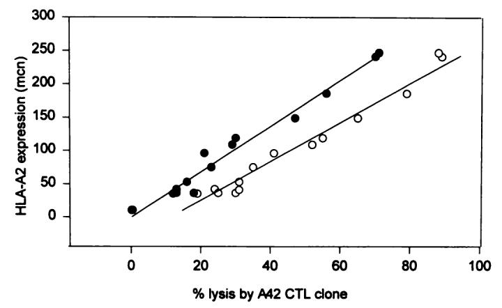
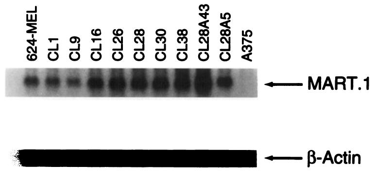
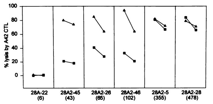
MHC class I antigen expression is necessary for CD8+ T-cell-mediated recognition of tumors. Recently, several mechanisms leading to loss or decreased expression of MHC antigens on the tumor cell surface have been described that may account for tumor escape from immune recognition. It is yet unknown whether tumor recognition by CTL occurs at a threshold amount of MHC molecules or correlates with the level of HLA-allele expression. In this study, a model was developed in which clones derived from the 624-MEL melanoma cell line and expressing varying amounts of HLA-A2 molecules were lysed in a standard 51Cr release assay by an HLA-A2-restricted CTL clone (A42) or a bulk culture of tumor-infiltrating lymphocytes. The A42 clone and the tumor-infiltrating lymphocyte culture were characterized previously as specifically recognizing the melanoma antigen MART-1(27-35) peptide. A marked heterogeneity in the susceptibility to lysis by A42 was observed in tumor clones and was not due to heterogeneous expression of MART-1 by the clones or loss of accessory molecules involved in the lymphocyte-target interaction. Lysis by A42 and by the tumor-infiltrating lymphocyte culture significantly correlated with the level of HLA-A2 expression, evaluated as mean channel number of fluorescence by flow cytometry (P < 0.001). Transfection of an HLA-A2-negative clone (624.28) with the HLA-A2.1 gene produced a panel of clones expressing different levels of HLA-A2, the lysis of which was highly correlated with the expression of HLA-A2 (P < 0.001). The addition of exogenous MART-1(27-35) peptide enhanced lysis of clones expressing intermediate amounts of HLA-A2 but did not affect clones with high expression. These data suggest that the number of HLA molecules present on the surface of tumor cells can quantitatively affect their lysis by CTL in situations with borderline amounts of peptide and/or MHC.
Figures
References
-
- Wolfel T, Klehmann E, Muller C, Schutt KH, Meyer zum Buschenfelde KH, Knuth A. Lysis of human melanoma cells by autologous cytolytic T cell clones: identification of human histocompatibility leukocyte antigen A2 as a restriction element for three different antigens. J. Exp. Med. 1989;170:797–810. - PMC - PubMed
-
- Crowley NJ, Darrow TL, Quinn-Allen MA, Seigler HF. MHC-restricted recognition of autologous melanoma by tumor-specific cytotoxic T cells: evidence for restriction by a dominant HLA-A allele. J. Immunol. 1991;146:1692–1699. - PubMed
-
- Rosenberg SA, Packard BS, Aebersold PM, et al. Use of tumor-infiltrating lymphocytes and interleukin-2 in the immunotherapy of patients with metastatic melanoma: a preliminary report [see comments] N. Engl. J. Med. 1988;319:1676–1680. - PubMed
-
- Pellegrino MA, Ferrone S, Reisfeld RA, Irie RF, Golub SH. Expression of histocompatibility (HLA) antigens on tumor cells and normal cells from patients with melanoma. Cancer (Phila.) 1977;40:36–41. - PubMed
-
- Ruiter DJ, Mattijssen V, Broecker EB, Ferrone S. MHC antigens in human melanomas. Semin. Cancer Biol. 1991;2(Suppl 1):35–45. - PubMed
MeSH terms
Substances
Grants and funding
LinkOut - more resources
Full Text Sources
Other Literature Sources
Medical
Research Materials