. 1995 Jul 14;82(1):101-10.
doi: 10.1016/0092-8674(95)90056-x.

Switches in expression of Plasmodium falciparum var genes correlate with changes in antigenic and cytoadherent phenotypes of infected erythrocytes

Affiliations

Switches in expression of Plasmodium falciparum var genes correlate with changes in antigenic and cytoadherent phenotypes of infected erythrocytes

J D Smith et al. Cell. .

Abstract

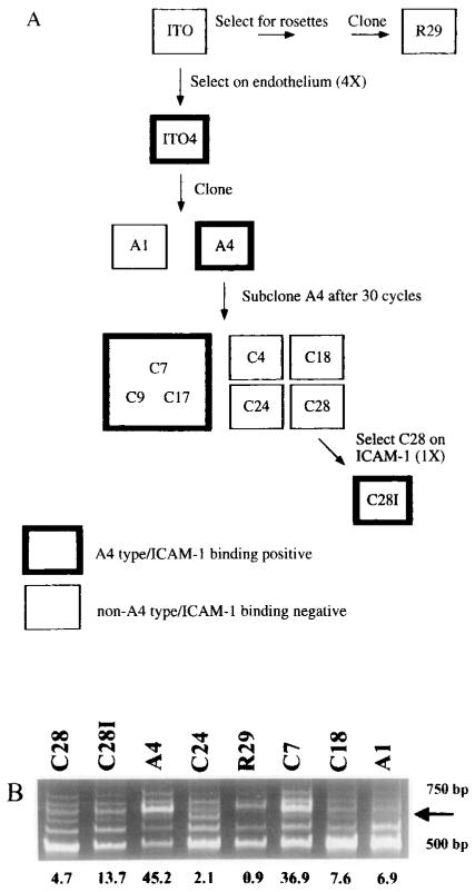
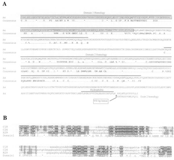
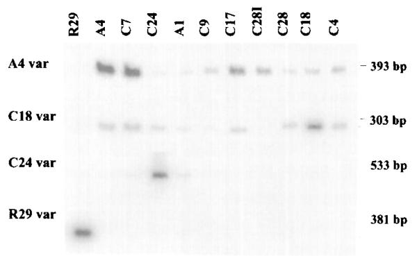
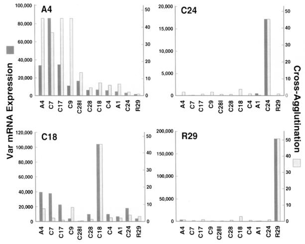
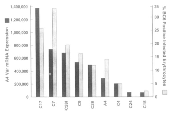
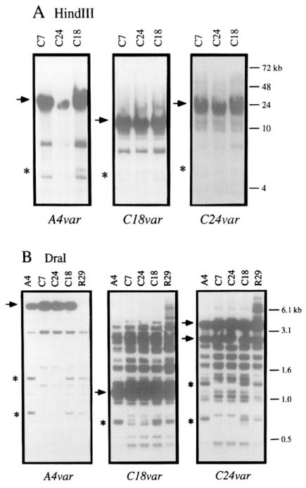
Plasmodium falciparum expresses on the host erythrocyte surface clonally variant antigens and ligands that mediate adherence to endothelial receptors. Both are central to pathogenesis, since they allow chronicity of infection and lead to concentration of infected erythrocytes in cerebral vessels. Here we show that expression of variant antigenic determinants is correlated with expression of individual members of a large, multigene family named var. Each var gene contains copies of a motif that has been previously shown to bind diverse host receptors; expression of a specific var gene correlated with binding to ICAM-1. Thus, our findings are consistent with the involvement of var genes in antigenic variation and binding to endothelium.


Figures

Figure 1
Figure 1
Degenerate Oligonucleotide Primers to DBL Domains Amplify a Message That Correlates with A4-Specific Agglutination of Members of the ITO Clonal Family (A) P. falciparum clonal tree. Clones in this family tree were derived as described previously by Roberts et al. (1992). Bold boxes indicate that the clone is of the A4 antigenic type and binds ICAM-1. Plain boxes indicate that the parasite is of a different antigenic type from A4 and other clones and that it does not bind ICAM-1. (B) RNA from antigenically similar or distinct clones was DNase treated, reverse transcribed, and amplified with the DBL primers UNIEBP5′ and UNIEBP3′. The arrow on the right side of the figure identifies a band whose intensity correlates with A4 antigenicity. Numbers shown below Figure 1B represent antigenic similarity to the A4 clone as defined by chance of heterologous cross-agglutination (see Experimental Procedures).
Figure 2
Figure 2
Deduced Amino Acid Sequence of var Genes Isolated from Antigenically Variant Clones (A) Alignment of the A4var sequence with the consensus sequence from the var genes cloned by Su et al. (1995). The deduced amino acid sequence encoded by A4var is compared with consensus sequence of the var DBL domains to which these genes are most similar (Su et al., 1995). Above the A4var sequence, a bold line indicates the regions of A4var homology with DBL domain 3 or DBL domain 2. At positions within the consensus sequence at which a number of similar amino acids can be found, the following symbols were used: U (I, L, V, M), at symbol (Y, F, W), ampersand (I, L, V, M, Y, F, W), percent (E, D), and dollar sign (S, T). The asterisk in the consensus sequence indicates gaps in the sequence where different numbers of amino acids are present. Boxed sequence identifies the original A4var sequence amplified with UNIEBP5′ and UNIEBP3′. A bold line, labeled hydrophobic, at the 3′ end of exon 1 indicates the putative transmembrane region. The position of a 950 bp intron is marked with an arrow, and the area of exon 2 homology is shown with consensus amino acid residues from other var sequences identified by Su et al. (1995). (B) The predicted amino acid sequences of C18var, C24var, and R29var genes are homologous with var DBL domain 1. Predicted amino acid sequences from the C18, C24, and R29 var genes were aligned with the consensus amino acid sequence of var DBL domain 1 defined by Su et al. (1995). The same symbols were used as above. Amino acids in the var sequences are highlighted with reverse print if a consensus amino acid is always present at that position in the var sequences identified by Su et al. (1995) and are highlighted in stippled print if a consensus amino acid is the most common of more than one amino acid present at that position.
Figure 3
Figure 3
Antigenically Distinct P. falciparum Clones Express Distinct var Messages RT–PCR using specific primers to var genes isolated from the A4, C18, C24, or R29 clones (indicated at the left of the figure) was performed on RNA isolated from each of the 11 clones listed at the top of the figure. RT–PCR products were hybridized with the respective var probes. The results are representative of three experiments (A4var) or two experiments (other var genes) performed on the same preparation of cDNAs.
Figure 4
Figure 4
Expression of Distinct var Transcripts Correlates with the Surface Antigenicty of P. falciparum–Infected Erythrocytes The results of RT–PCR experiments with different var-specific primers on the panel of clones (Figure 3) were quantified by densitometry and compared with the cross-agglutination of each clone with either the A4, C24, C18, or R29 clone (indicated at the top left of each bar graph). The geometric means of RT–PCR products from three experiments using A4var primers and two experiments using C24var, C18var, or R29var primers are plotted next to cross-agglutination of each clone with A4, C18, C24, or R29 clones (see Experimental Procedures).
Figure 5
Figure 5
A4var Expression Correlates with the Percentage of P. falciparum-Infected Erythrocytes Binding a Monoclonal Antibody (MAb BC6) That Is Specific for A4 Variants The percentage of parasitized erythrocytes of the A4 type in different clones was determined by flow cytometry using MAb BC6. At the same time, RNA was extracted, and the level of A4var expression was determined by RT–PCR. The geometric mean of two RT–PCR experiments is plotted next to the percentage of A4-type variants in each clonal culture.
Figure 6
Figure 6
Southern Blot Analysis of var Gene Organization in Clones That Have Switched Antigenic Type (A) DNA from the C7, C24, and C18 clones was digested with HindIII and separated on a PFG. (B) DNA from the A4, C7, C18, C24, and R29 clones was digested with Dral and separated by standard electrophoresis. The probes used were A4var, C24var, or C18var as indicated. Major bands of hybridization are indicated with an arrow. The R29 clone has deleted the major A4var band of hybridization. Minor bands that are deleted are indicated by an asterisk.

Comment in

  • Antigenic variation in malaria.
    Borst P, Bitter W, McCulloch R, Van Leeuwen F, Rudenko G. Borst P, et al. Cell. 1995 Jul 14;82(1):1-4. doi: 10.1016/0092-8674(95)90044-6. Cell. 1995. PMID: 7606774 Review. No abstract available.

References

    1. Adams JA, Hudson DE, Torii M, Ward GE, Wellems TE, Aikawa M, Miller LH. The Duffy receptor family of Plasmodium knowlesi is located within the micronemes of invasive malaria merozoites. Cell. 1990;63:141–153. - PubMed
    1. Adams JH, Sim BKL, Dolan SA, Fang X, Kaslow DC, Miller LH. A family of erythrocyte binding proteins of malaria parasites. Proc. Natl. Acad. Sci. USA. 1992;89:7085–7089. - PMC - PubMed
    1. Barnwell JW, Asch AS, Nachman RL, Yamaya M, Aikawa M, Ingravallo P. A human 88-kD membrane glycoprotein (CD36) functions in vitro as a receptor for a cytoadherence ligand on Plasmodium falciparum-infected erythrocytes. J. Clin. Invest. 1989;84:765–772. - PMC - PubMed
    1. Baruch DI, Pasloske BL, Singh HB, Xiahui B, Ma XC, Feldman M, Taraschi TF, Howard RJ. Cloning the Plasmodium falciparum gene encoding PfEMP1, a malarial variant antigen and adherence receptor on the surface of parasitized human erythrocytes. Cell. 1995;82(this issue) - PubMed
    1. Berendt AR, Simmons DL, Tansey J, Newbold CI, Marsh K. Intercellular adhesion molecule-1 is an endothelial adhesion receptor for Plasmodium falciparum. Nature. 1989;341:57–59. - PubMed

Publication types

MeSH terms