. 1995 May 1;355(2):296-315.
doi: 10.1002/cne.903550208.

Characterization of inducible cyclooxygenase in rat brain

Affiliations

Characterization of inducible cyclooxygenase in rat brain

C D Breder et al. J Comp Neurol. .

Abstract

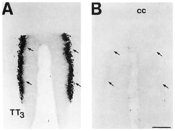
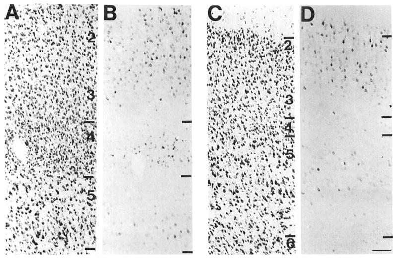
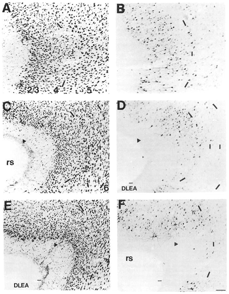
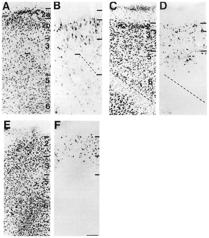
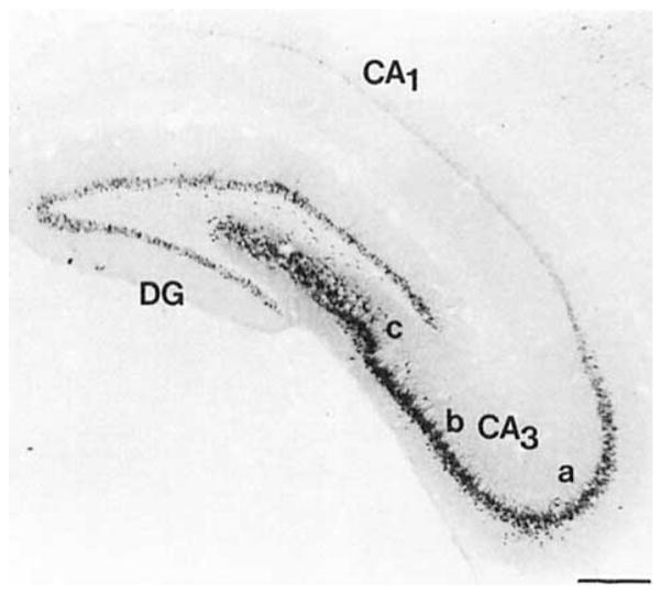
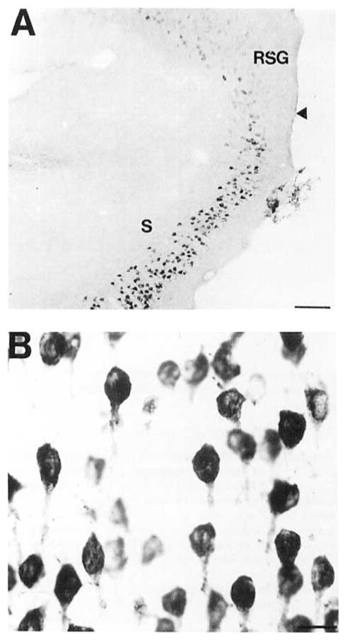
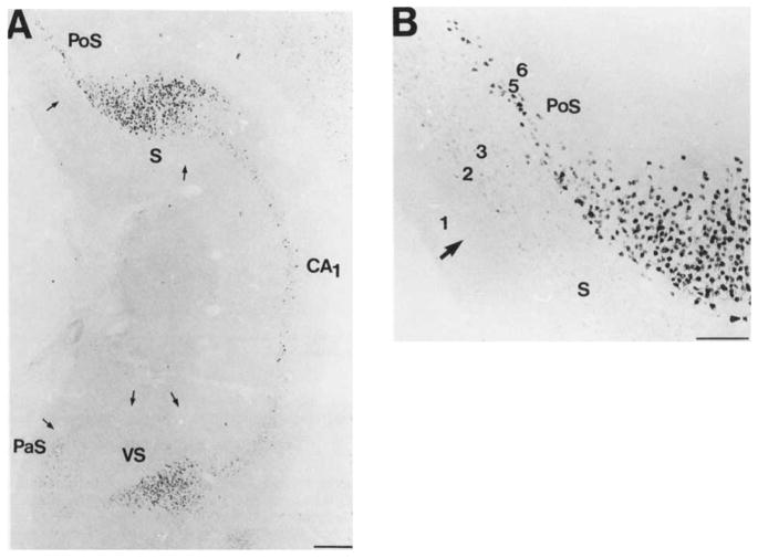
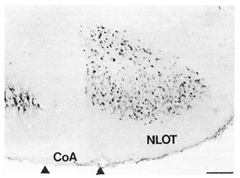
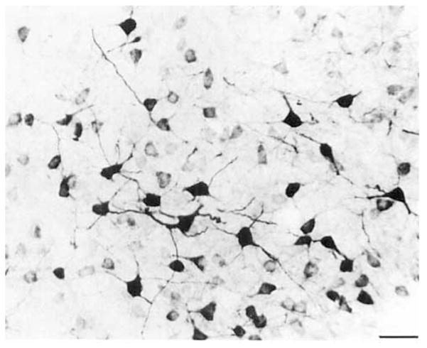
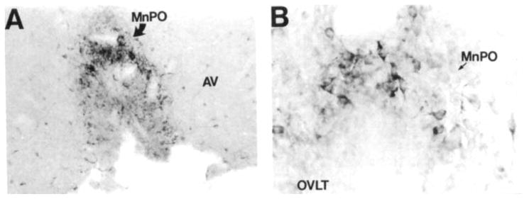
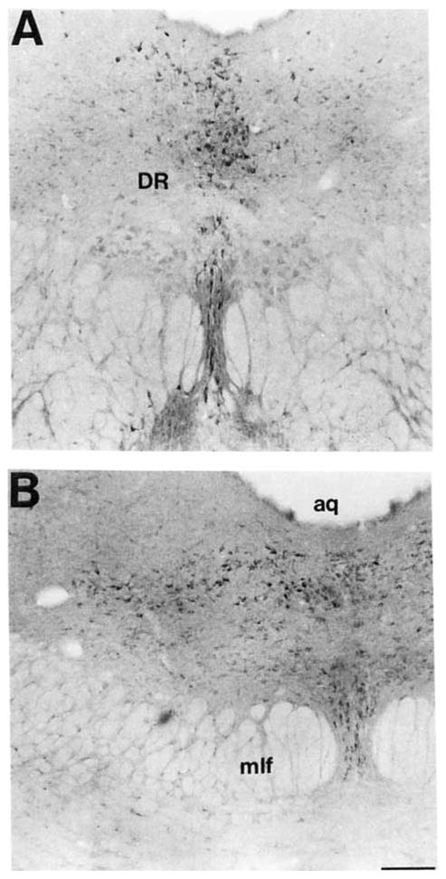
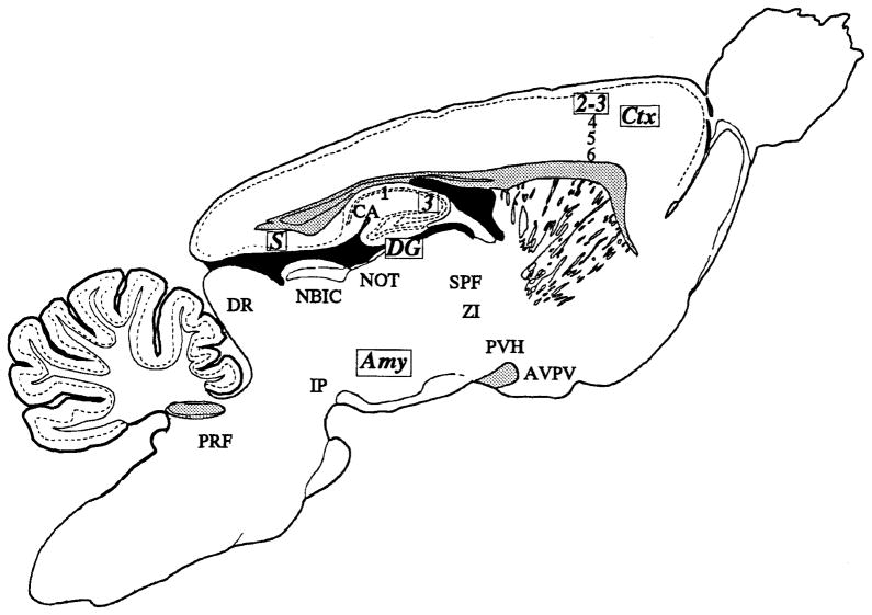
Considerable debate exists regarding the cellular source of prostaglandins in the mammalian central nervous system (CNS). At least two forms of prostaglandin endoperoxide synthase, or cyclooxygenase (COX), the principal enzyme in the biosynthesis of these mediators, are known to exist. Both forms have been identified in the CNS, but only the distribution of COX 1 has been mapped in detail. In this study, we used Western blot analysis and immunohistochemistry to describe the biochemical characterization and anatomical distribution of the second, mitogen-inducible form of this enzyme, COX 2 in the rat brain. COX 2-like immunoreactive (COX 2-ir) staining occurred in dendrites and cell bodies of neurons, structures that are typically postsynaptic. It was noted in distinct portions of specific cortical laminae and subcortical nuclei. The distribution in the CNS was quite different from COX 1. COX 2-ir neurons were primarily observed in the cortex and allocortical structures, such as the hippocampal formation and amygdala. Within the amygdala, neurons were primarily observed in the caudal and posterior part of the deep and cortical nuclei. In the diencephalon, COX 2-ir cells were also observed in the paraventricular nucleus of the hypothalamus and in the nuclei of the anteroventral region surrounding the third ventricle, including the vascular organ of the lamina terminalis. COX 2-ir neurons were also observed in the subparafascicular nucleus, the medial zona incerta, and pretectal area. In the brainstem, COX 2-ir neurons were observed in the dorsal raphe nucleus, the nucleus of the brachium of the inferior colliculus, and in the region of the subcoeruleus. The distribution of COX 2-ir neurons in the CNS suggests that COX 2 may be involved in processing and integration of visceral and special sensory input and in elaboration of the autonomic, endocrine, and behavioral responses.


Figures

Fig. 1
Fig. 1
Western blot analysis of cyclooxygenase 2-like immunoreactivity (COX 2-ir). Lanes: A: rat brain cerebral cortex; B: rat brain cerebellum; C: lipopolysaccharide (LPS)-stimulated RAW 243.7 cells; and D: unstimulated RAW 243.7 cells. The arrows denote the position of the COX 2 protein.
Fig. 2
Fig. 2
A: Brightfield photomicrograph of COX 2-ir neurons in the granular cell layer of the dorsal, TT3, region of the tenia tecta. B: The adjacent section stained with the primary COX 2-ir antiserum pread- sorbed with the DD-2 peptide. Note the lack of staining despite intensification of the control material. The arrows denote the position of the granular cell layer of this division. Scale bar = 150 μm.
Fig. 3
Fig. 3
A series of brightfield photomicrographs demonstrate the laminar organization of COX 2-ir neurons in motor fields of the cortex (A,C). Laminae are indicated on the adjacent Nissl-stained sections (B,D). A,B: Primary motor cortex. C,D: Frontal eye field. Scale bar = 100 μm.
Fig. 4
Fig. 4
A series of brightfield photomicrographs demonstrate the laminar organization of COX 2-ir neurons in somatosensory fields of the cortex (A,C). Laminae are indicated on the adjacent Nissl-stained sections (B,D). A,B: Primary somatosensory cortex. C,D: Secondary somatosensory cortex. Scale bar = 100 μm
Fig. 5
Fig. 5
A series of brightfield photomicrographs demonstrate the laminar organization of COX 2-ir neurons in several levels of the perirhinal cortex (A,C,E). Photomicrographs are oriented in the normal transverse view. The arrowheads denote the deepest portion of the invagination of the rhinal sulcus, which we have taken as the division of the dorsal and ventral portions of this cortical field. Laminae are indicated in the adjacent Nissl-stained sections (B,D,F). A,B: Rostral perirhinal cortex, approximately at the level of Figure 19 of Zilles (1985). C,D: Intermediate perirhinal cortex, approximately at the level of Figure 23 of Zilles (1985). E,F: Caudal perirhinal cortex, approximately at the level of Figure 25 of Zilles (1985). Dashes in A, C, and E correspond to the lamina boundaries in B, D, and F. Scale bar = 100 μm.
Fig. 6
Fig. 6
A series of brightfield photomicrographs demonstrate the laminar organization of COX 2-ir neurons in different fields of the entorhinal cortex (A,C,E). The dorsal aspect (closest to the rhinal sulcus) of each section is positioned on the right side. Laminae are indicated in the adjacent Nissl-stained sections (B,D,F) and are further delineated in certain sections by the use of dashed lines. A,B: Lateral entorhinal cortex. C,D: Ventrolateral entorhinal cortex. E,F: Medial entorhinal cortex. Scale bar = 100 μm.
Fig. 7
Fig. 7
A low-power brightfield photomicrograph of COX 2-ir neurons in the dorsal hippocampal formation. Scale bar = 300 μm.
Fig. 8
Fig. 8
A pair of brightfield photomicrographs show COX 2-ir neurons in the cell fields of the Cornu ammonis. A: COX 2-ir neurons in the C& field. Note the dense staining of cells in the stratum pyramidalis and sparse population of COX 2-ir cells in the stratum radiatum. B: A sparse population of COX 2-ir neurons were observed in the stratum pyramidalis of the CA1 field. Scale bar = 50 μm.
Fig. 9
Fig. 9
A pair of brightfield photomicrographs depict COX 2-ir neurons in the dorsal subiculum. A: A low-power photomicrograph of COX 2-ir neurons in the subiculum at the level of the splenium of the corpus callosum. The arrowhead denotes the border with the retrosplenial granular cortex. B: A high-power photomicrograph of the COX 2-ir neurons in the dorsal subiculum. Note the uniformity of the ventral processes projecting from these neurons. Scale bars = 100 μm in A, 20 μm in B.
Fig. 10
Fig. 10
A pair of brightfield photomicrographs depict the COX 2-ir neurons in the temporal portion of the hippocampal formation. A: A low-power photomicrograph depicts COX 2-ir neurons in the dorsal and ventral portions of the subiculum, postsubiculum, and stratum pyramidal of CA1. The arrows delineate regional boundaries in the subicular region of the hippocampal formation. B: High-power photomicrograph depicts the COX 2-ir neurons in the dorsal division of the temporal part of the subiculum and the laminar organization of the COX 2-ir neurons in the postsubiculum. The numbers denote the laminae in the postsubiculum and the arrow marks its border with the dorsal subiculum. Scale bars = 300 μm in A, 150 μm in B.
Fig. 11
Fig. 11
A pair of low-power brightfield photomicrographs depict the COX 2-ir neurons in the (A)middle level of the amygdala and (B) posterior level of the amygdala. Scale bar = 300 μm.
Fig. 12
Fig. 12
A hrightfield photomicrograph depicts COX 2-ir neurons in the nucleus of the lateral olfactory tract. Scale bar = 200 μm.
Fig. 13
Fig. 13
A high-power photomicrograph depicts COX 2-ir neurons in the lateral nucleus of the amygdala. Scale bar = 30 μm.
Fig. 14
Fig. 14
A series of brightfield photomicrographs depict COX 2-ir neurons in the anteroventral region of the third ventricle. A: A low-power photomicrograph depicts COX 2-ir neurons in the rostra1 anteroventral region of the third ventricle. Dense aggregates of these neurons were observed in the median preoptic nucleus and vascular organ of the lamina terminalis and less densely in the anteroventral periventricular nucleus. B: A high-power photomicrograph depicts COX 2-ir neurons in the dorsal aspect of the third ventricle in the median preoptic nucleus. Scale bars = 100 μm in A, 30 μm in B.
Fig. 15
Fig. 15
A pair of brightfield photomicrographs depict COX 2-ir neurons in the paraventricular nucleus of the hypothalamus. A: A low-power photomicrograph depicts the COX 2-ir neurons in the ventral portion of the medial parvocellular subnucleus of the paraventricular nucleus of the hypothalamus. B: A high-power photomicrograph depicts the COX 2-ir neurons in this division. Scale bars = 150 μm in A, 30 μm in B.
Fig. 16
Fig. 16
A brightfield photomicrograph shows COX 2-ir neurons ventromedial to the mammillothalamic tract in the zona incerta and dorsal hypothalamic area. Scale bar = 150 μm.
Fig. 17
Fig. 17
A pair of hrightfield photomicrographs depict the COX 2-ir neurons in the medial thalamus. A: At intermediate levels of this group, the cells outlined the magnocellular division of the subparafascicular nucleus. B: Caudally, this group extended dorsally through the diencephalic central gray. Scale bar = 150 μm.
Fig. 18
Fig. 18
A low-power photomicrograph shows COX 2-ir neurons in the interpeduncular nucleus. Within this structure, these cells were only observed in the dorsal lateral subnucleus. Scale bar = 150 μm.
Fig. 19
Fig. 19
A pair of brightfield photomicrographs depict COX 2-ir neurons in the dorsal raphe nucleus. A: A brightfield photomicrograph depicts COX 2-ir neurons in the rostral dorsal and ventral portions of the dorsal raphe nucleus. B: A brightfield photomicrograph depicts COX 2-ir neurons in the caudal dorsal, ventral, and lateral portions of the dorsal raphe nucleus. Scale bar = 150 μm.
Fig. 20
Fig. 20
Schematic diagram shows the distribution of COX 2-ir neurons in the rat brain. Regions noted in bold and italic type with boxed abbreviations had the densest and most intensely staining COX 2-ir neurons. Other regions noted here had more modest numbers of relatively lighter stained COX 2-ir neurons. Stippled regions represent major fiber tracts in the central nervous system (CNS) and are meant to serve as anatomical landmarks.

References

    1. Ben-Ari Y, Le Gal LaSalle G. Lateral amygdala unit activity: II. Habituating and non-habituating neurons. Electroenceph Clin Neuro-physiol. 1974;37:463–472. - PubMed
    1. Bentivoglio M, Molinari M. The interrelationships between cell groups in the caudal diencephalon of the rat to the striatum and to the medulla oblongota. Exp Brain Res. 1984;54:57–65. - PubMed
    1. Berkley KJ, Mash DC. Somatic sensory projections to the pretectum in the cat. Brain Res. 1978;158:445–449. - PubMed
    1. Berkley KJ, Budell RJ, Blomqvist A, Bull M. Output systems of the dorsal column nuclei in the cat. Brain Res Rev. 1986;11:199–225. - PubMed
    1. Berson DM, Graybiel AM. Some cortical and subcortical fiber projections to the accessory optic nuclei in the cat. Neuroscience. 1980;5:2203–2217. - PubMed

Publication types

Substances