Review
. 1995 Aug;26(8):816-23.
doi: 10.1016/0046-8177(95)90001-2.

Glial cytokines in Alzheimer's disease: review and pathogenic implications

Affiliations
Review

Glial cytokines in Alzheimer's disease: review and pathogenic implications

R E Mrak et al. Hum Pathol. 1995 Aug.

Abstract

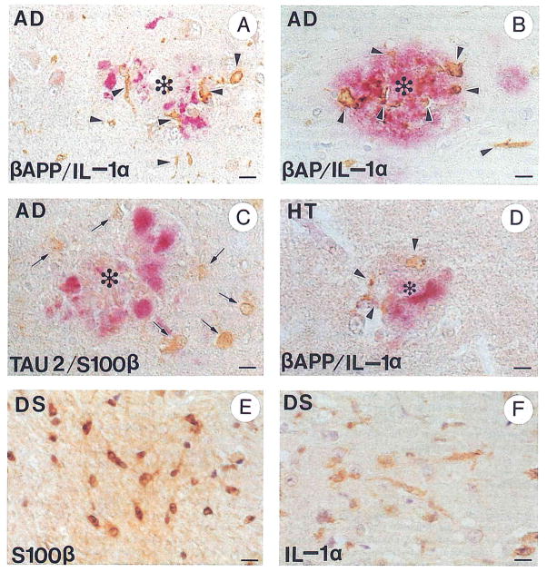
The roles of activated glia and of glial cytokines in the pathogenesis of Alzheimer's disease are reviewed. Interleukin-1 (IL-1), a microglia-derived acute phase cytokine, activates astrocytes and induces expression of the astrocyte-derived cytokine, S100 beta, which stimulates neurite growth (and thus has been implicated in neuritic plaque formation) and increases intracellular free calcium levels. Interleukin-1 also upregulates expression and processing of beta-amyloid precursor proteins (beta-APPs) (thus favoring beta-amyloid deposition) and induces expression of alpha 1-antichymotrypsin, thromboplastin, the complement protein C3, and apolipoprotein E, all of which are present in neuritic plaques. These cytokines, and the molecular and cellular events that they engender, form a complex of interactions that may be capable of self-propagation, leading to chronic overexpression of glial cytokines with neurodegenerative consequences. Self-propagation may be facilitated by means of several reinforcing feedback loops. beta-Amyloid, for instance, directly activates microglia, thus inducing further IL-1 production, and activates the complement system, which also leads to microglial activation with IL-1 expression. Self-propagation also could result when S100 beta-induced increases in intraneuronal free calcium levels lead to neuronal injury and death with consequent microglial activation. Such chronic, self-propagating, cytokine-mediated molecular and cellular reactions would explain the progressive neurodegeneration and dementia of Alzheimer's disease.


Figures

FIGURE 1
FIGURE 1
Immunohistochemical demonstration of IL-1α+ activated microglia and S100β+ activated astrocytes in Alzheimer’s disease and Down’s syndrome, and following head trauma. (A) Alzheimer’s disease: activated IL-1α+ microglia (brown, arrowheads) associated with β-APP+ dystrophic neurites (red) in a neuritic plaque (*). (B) Alzheimer’s disease: activated IL-1α+ microglia (brown, arrowheads) associated with β-amyloid (β-AP) immunopositive deposits (red) in a neuritic plaque (*). (C) Alzheimer’s disease: activated S100β+ astrocytes (brown, arrows) associated with Tau 2+ dystrophic neurites (red) in a neuritic plaque (*). (D) Head trauma: activated IL-1α+ microglia (brown, arrowheads) associated with β-APP+ dystrophic neurites in a neuritic plaque-like structure in a patient surviving 48 hours after head trauma. (E) Down’s syndrome: activated S100β+ astrocytes (brown) in a trisomy 21 fetus at 23 weeks’ gestation; (F) Down’s syndrome: activated IL-1α+ microglia in a 2-year-old patient with Down’s syndrome. Bars represent 15 μm. Immunohistochemical technique: Paraffin blocks of formalin-fixed mesial temporal lobe tissue were sectioned at 10-μm thickness. Sections were deparaffinized in xylene, rehydrated in a graded series of ethanol solutions, and permeabilized in 0.05% Triton-X 100 for 10 minutes followed by 0.2 N HCl for 20 minutes. Endogenous peroxidase was blocked by incubation with 3% H2O2 in 97% methanol for 30 minutes. Sections for β-APP immunoreaction were pretreated with 99% formic acid for 5 minutes and washed with phosphate-buffered saline. Antibodies employed were polyclonal anti-IL-1α (Cistron, Pine Brook, NJ), diluted 1:20; polyclonal anti-β-amyloid (Boehringer-Mannheim Biochemica, Indianapolis, IN), diluted 1:10; monoclonal anti-β-amyloid (a gift from G.W. Roberts, SmithKline Beecham Pharmaceuticals, Essex, UK), diluted 1:1,000; monoclonal anti-β-APP (clone 22C11; Boehringer-Mannheim Biochemica), diluted 1:10; polyclonal antibovine S100β (a gift from L.J. Van Eldik, Northwestern University, Chicago, IL), diluted 1:300; and monoclonal Tau 2 antibody (Sigma Chemical Company, St Louis, MO), diluted 1:100. For dual immunoreaction sections were further processed according to the manufacturer’s protocol in DAKO’s (Glostrup, Denmark) double immunolabeling kit (K-665).
FIGURE 2
FIGURE 2
Diagram of proposed pathogenic interactions between glial cytokines and molecular and cellular events in Alzheimer’s disease. Micro, microglial; Astro, astroglial; PN-1, protease nexin 1; α1-ACT, α1 antichymotrypsin; (Ca)i, intracellular free calcium concentration; P-tau, excessively phosphorylated tau; PHF, paired helical filaments in neurofibrillary tangles; PolyA+ mRNA, polyadenylated mRNA.

References

    1. Selkoe DJ. Alzheimer’s disease: A central role for amyloid. J Neuropathol Exp Neurol. 1994;53:438–447. - PubMed
    1. Davies CA, Mann DMA, Sumpter PQ, et al. A quantitative morphometric analysis of the neuronal and synaptic content of the frontal and temporal cortex in patients with Alzheimer’s disease. J Neurol Sci. 1987;78:151–164. - PubMed
    1. Terry RD, Masliah E, Salmon DP, et al. Physical basis of cognitive alterations in Alzheimer’s disease: Synapse loss is the major correlate of cognitive impairment. Ann Neurol. 1991;30:572–580. - PubMed
    1. Masliah E, Terry RD, Alford M, et al. Cortical and subcortical patterns of synatophysin-like immunoreactivity in Alzheimer’s disease. Am J Pathol. 1991;138:235–246. - PMC - PubMed
    1. DeKosky ST, Scheff SW. Synapse loss in frontal cortex biopsies in Alzheimer’s disease: Correlation with cognitive severity. Ann Neurol. 1990;27:457–464. - PubMed

Publication types