Comparative Study
. 1995 Aug 25;82(4):599-609.
doi: 10.1016/0092-8674(95)90032-2.

Localization of centromere function in a Drosophila minichromosome

Affiliations
Comparative Study

Localization of centromere function in a Drosophila minichromosome

T D Murphy et al. Cell. .

Abstract

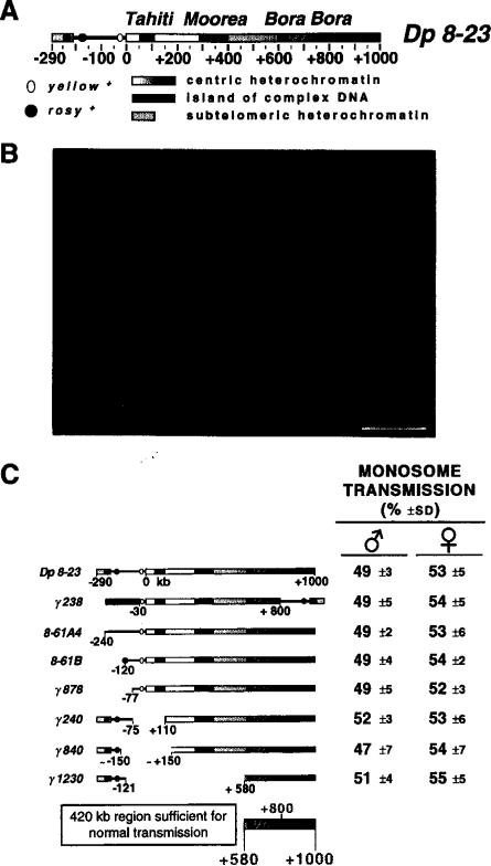
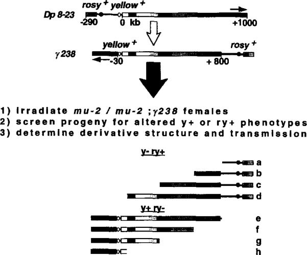
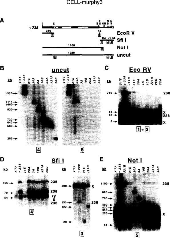
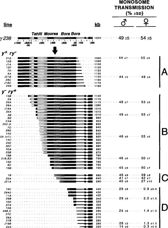
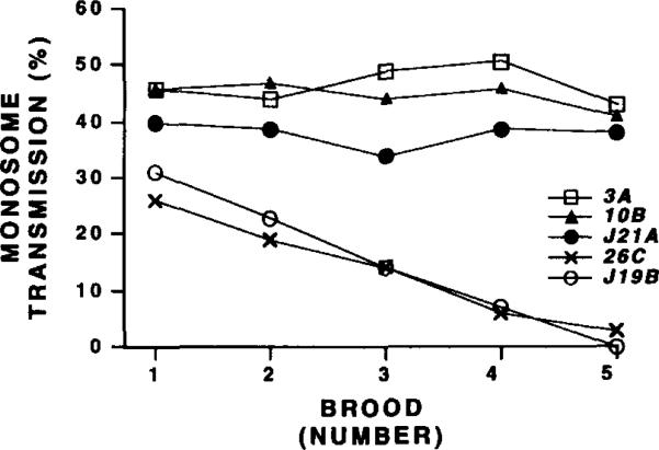
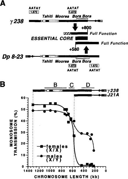
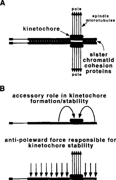
The DNA elements responsible for centromere activity in a metazoan have been localized using the Drosophila minichromosome Dp1187. Deleted minichromosomes were generated by irradiation mutagenesis, and their molecular structures were determined by pulsed-field Southern blot analysis. Analyses of the transmission behavior of Dp1187 derivatives localized sequences necessary for chromosome inheritance within the centric heterochromatin. The essential core of the centromere is contained within a 220 kb region that includes significant amounts of complex DNA. Completely normal inheritance also requires approximately 200 kb on either side of the essential core. This flanking DNA predominantly contains highly repeated sequences, and the amount required for normal transmission differs among division types and between the sexes. We propose that the essential core is the site of kinetochore formation and that flanking DNA provides two functions: sister chromatid cohesion and indirect assistance in kinetochore formation or function.


Figures

Figure 1
Figure 1. The Structure of Dp1187 and Gross Localization of a Centromere
(A) Structure of Dp 8-23. Dp 8-23 contains sequences from the X-tip euchromatin (black line) and subtelomeric heterochromatin (closed box) juxtaposed to 1 Mb of X centric heterochromatin (stippled gradient, 0 to +1000 kb). Two ry+ marker genes (stippled circles) introduced by P element transposition are present in the Dp1187 derivative Dp 8-23 (Tower et al., 1993), in addition to the y+ gene (open oval) normally present in the X and Dp1187 euchromatin. The kilobase coordinates correspond to those determined for the parental Dp1187 (Karpen and Spradling, 1990, 1992); the actual size of Dp 8-23 is 1320 kb, because each PZ insertion adds 14.5 kb to the 1290 kb Dp1187 length. The centric heterochromatin contains islands of complex DNA (single-copy or middle-repetitive sequences; closed boxes, Tahiti, Moorea, and Bora Bora) separated by blocks composed predominantly of highly repeated satellite DNA (stippled blocks). At least eight enzymes failed to cut in the central region of Bora Bora (diagonal bars); this region may contain satellite DNA or an unusual chromatin structure (Le et al., 1995). (B) Cytological visualization of Dp8-23. A metaphase squash from a third instar larval brain neuroblast is shown (bar equals 5 μm). The monosomic minichromosome (Dp) and the X, 2, 3, and 4 chromosome pairs are marked. (C) Gross localization of a Dp1187 centromere. The monosome transmission behavior of Dp 8-23 derivatives was monitored from both male and female parents (percent ± SD; see Experimental Procedures). From these analyses we conclude that a centromere and other sequences essential to inheritance through mitosis and meiosis are located maximally between +580 and +1000, which includes Bora Bora and satellite DNA.
Figure 2
Figure 2. Generating Bidirectional Deletion Derivatives Using the γ238 Inversion
The structures of Dp 8-23 and the irradiation-induced derivative γ238 are shown (Le et al., 1995; orientation of the centric heterochromatin is indicated by the stippled gradient and arrows). Terminal deficiencies were generated and analyzed as described in steps 1, 2, and 3 (see Experimental Procedures). The mutation mu2 allows terminal deficiencies to be recovered at very high frequencies after females are treated with low radiation doses (Mason et al., 1984). y- ry+ lines are candidates for harboring minichromosomes with deletions in the left end of γ238, while y+ ry- lines could contain minichromosomes that lacked the right end. Hypothetically, random induction of terminal breaks during γ238 mutagenesis could generate derivatives with the structures shown below (a–h).
Figure 3
Figure 3. Structural Analyses of γ238 Derivatives
(A) Restriction fragments used to analyze the structures of γ238 derivatives. The relevant restriction sites are shown above the chromosome (E, EcoRV; S, Sfil; N, Notl), while the fragments are shown as thick closed lines below. The sizes of the fragments in kilobases are indicated above each line, while the probes (1–6, in boxes) are indicated below at their chromosomal locations (see Experimental Procedures). (B–E) Examples of pulsed-field Southern blot hybridization analyses performed on uncut, EcoRV-digested, Sfil-digested, and Notl-digested high molecular weight DNAs. See Experimental Procedures for further analyses of the results. Sizes of bands are shown on the left of each blot (in kilobases), and the genomic sources of fragments present in the parental genotype (y; ry506; γ238) are indicated on the right (238, γ238; X, endogenous X chromosome; ry, endogenous ry locus). The probes hybridized to each blot are indicated below. J21B and 31E were recovered as y+ ry- lines, while the other derivatives shown in this figure were y- ry+ lines. Minichromosomes and restriction fragments that contain large amounts of heterochromatin displayed weak signals (e.g., 210 kb fragment in EcoRV digest and uncut chromosomes >1000 kb), likely owing to incomplete transfer to membranes (Glaser and Spradling, 1994) and inefficient probe hybridization. The following run conditions were used: (B) uncut DNA, 50–110 s, 2 s ramp, 28 hr at 180V, 160 mA; (C) EcoRV digests, 1–30 s, 1 s ramp, 18 hr at 180V, 160 mA; (D) Sfil digests, 1–30 s, 1 s ramp, 24 hr at 180V, 160 mA; (E) Notl digests, 40–100 s, 2 s ramp, 22 hr at 180V, 160 mA.
Figure 4
Figure 4. Structures and Transmission Behavior of γ238 Derivatives
The structures of the γ238 derivatives (line designations on the left) were determined by pulsed-field Southern blot analyses (Figure 3; see Experimental Procedures). The total size is displayed to the right of each structure in kilobases and ranged from 225 kb to 1190 kb. The monosome transmission behavior of selected derivatives was determined as before (see Figure 1C and Experimental Procedures). Groups A and B displayed normal transmission in males and females, group C was partially unstable in females, and group D was highly unstable in females and partially unstable in males.
Figure 5
Figure 5. Transmission of Acentric γ238 Derivatives Decreases During Brooding
The monosome transmission (Y-axis, percent) of five derivatives (3A, 10B, J21A, 26C, and J19B) was determined for males mated to fresh virgin females at 2, 5, 8, 11, and 14 days after eclosion (X-axis; broods 1-5, respectively). Only chromosomes that lack Bora Bora (26C and J19B) displayed significantly reduced transmission during brooding.
Figure 6
Figure 6. Summary of the Regions Required for Dp1187 Transmission
(A) Localizing the Dp1187 centromere. Summary of the results from the transmission tests of the Dp 8-23 and γ238 derivatives. For the γ238 derivatives (Figures 4 and 5), the +510 to +800 region (stippled box) promoted moderate stability, whereas another 140 kb of centric heterochromatin was necessary for full function (stippled diagonals). The sufficiency of the +580 to +1000 region (lower stippled box) was demonstrated with the Dp 8-23 derivatives (Figure 1C). Thus, the +580 to +800 region of overlap (closed box, Bora Bora) is essential to achieve stable transmission, but 200 kb of flanking heterochromatin on either side of Bora Bora provides a redundant function necessary for completely normal behavior. The left and right flanking regions both contain blocks of the 1.672 (AATAT) satellite DNA (Le et al., 1995). (B) Comparison of the transmission behavior of γ238 derivatives in males and females. The monosome transmission of y- ry+ derivatives (Y-axis, percent) is plotted relative to length (X-axis, kilobases). Data taken from Figure 4. Note that the shape of the two curves is similar, but transmission in males is affected less by deletion of flanking heterochromatin or the removal of Bora Bora. The breakpoint distributions for groups B, C, and D (Figure 4) are indicated.
Figure 7
Figure 7. Models for Transmission Functions Encoded by Dp1187 Centric Heterochromatin
The Bora Bora essential core is proposed to encode kinetochore formation (large stippled ovals), which interacts with spindle microtubules (closed arrows) to promote chromosome movement. (A) Here the flanking heterochromatin is proposed to play a role in sister chromatid cohesion. Cytological analyses suggest that many different heterochromatic sequences can participate in this redundant function (light stippled circles), but our results define the minimal DNA sequences to be 200 kb on either side of Bora Bora (dark stippled circles). (B) The flanking heterochromatin also could play an accessory role in kinetochore formation, stability, or both. In the model displayed on top, the flanking regions act in cis to assist kinetochore function at Bora Bora, for example by altering chromatin structure. An alternative model is displayed below, in which antipoleward microtubule-mediated forces (Rieder and Salmon, 1994) act on the flanking heterochromatin. Such forces would indirectly promote kinetochore stability at Bora Bora by creating tension (Li and Nicklas, 1995; Murphy and Karpen, 1995). Contrapoleward or interchromosomal forces are not shown, but could produce the same result. Again, 200 kb of flanking DNA would be sufficient for full function (dark stippled arrows), but this does not exclude the participation of other heterochromatic regions (light stippled arrows). In all three models, asingle chromosome in mitotic metaphase or meiosis II is shown, but the same functions could act on bivalents in meiosis I.

References

    1. Afshar K, Barton NR, Hawley RS, Goldstein LSB. DNA binding and meiotic chromosomal localization of the Drosophila nod kinesin-like protein. Cell. 1995;81:129–138. - PubMed
    1. Allshire RC, Cranston G, Gosden JR, Maule JC, Hastie ND, Fantes PA. A fission yeast chromosome can replicate autonomously in mouse cells. Cell. 1987;50:391–403. - PubMed
    1. Allshire RC, Javerzat JP, Redhead NJ, Cranston G. Position effect variegation at fission yeast centromeres. Cell. 1994;76:157–169. - PubMed
    1. Ashburner M. Drosophila: A Laboratory Handbook. Cold Spring Harbor Press; Cold Spring Harbor, New York: 1990.
    1. Ault JG, Rieder CL. Centrosome and kinetochore movement during mitosis. Curr. Opin. Cell Biol. 1994;6:41–49. - PubMed

Publication types

LinkOut - more resources