

doi: 10.1126/science.266.5189.1399.
Long-term behavioral recovery in parkinsonian rats by an HSV vector expressing tyrosine hydroxylase
Affiliations
- PMID: 7669103
- PMCID: PMC2638002
- DOI: 10.1126/science.266.5189.1399
Item in Clipboard
Long-term behavioral recovery in parkinsonian rats by an HSV vector expressing tyrosine hydroxylase
Science.
.
Abstract
One therapeutic approach to treating Parkinson's disease is to convert endogenous striatal cells into levo-3,4-dihydroxyphenylalanine (L-dopa)-producing cells. A defective herpes simplex virus type 1 vector expressing human tyrosine hydroxylase was delivered into the partially denervated striatum of 6-hydroxydopamine-lesioned rats, used as a model of Parkinson's disease. Efficient behavioral and biochemical recovery was maintained for 1 year after gene transfer. Biochemical recovery included increases in both striatal tyrosine hydroxylase enzyme activity and in extracellular dopamine concentrations. Persistence of human tyrosine hydroxylase was revealed by expression of RNA and immunoreactivity.
Figures
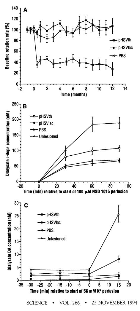
Rotation rates and striatal l -dopa or DA concentrations after stereotactic injection of pHSVth, pSHVlac, or PBS into the partially denervated striatum (). (A) The rats were tested at various times () for the apomorphine-induced rotation rate, and the values shown are the average percent of the baseline rotation rate for each group. (B) Striatal l -dopa concentrations were measured by microdialysis () after perfusion with NSD 1015 as an indication of striatal TH activity (). (C) Striatal DA levels were measured by microdialysis in low K+ (3 mM) and after perfusion with high K+ (56 mM) ().
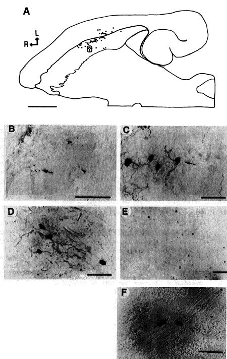
TH immunoreactivity was detected with an antibody to TH (78), and β-Gal was detected with X-Gal (). (A) through (C) show rat pHSVth no. 27. (A) Composite drawing of charted sections, showing the positions of 48 cells containing TH immunoreactivity in the striatum and neocortex. Every third section was analyzed. L, lateral; R, rostral; scale bar, 2 mm. (B) Low-magnification photomicrograph of clusters of striatal cells containing TH immunoreactivity. Arrowheads point to two clusters, and the arrow indicates a third cluster [boxed in (A)]; scale bar, 500 μm. (C) High-magnification view of a cluster of striatal cells containing TH immunoreactivity with neuronal morphology [boxed (A)]; scale bar, 50 μm. (D) and (E) show rat pHSVth no. 31. (D) A cluster of pallidal neurons containing TH immunoreactivity; scale bar, 50 μm (E) A cluster of cortical neurons (agranular frontal cortex, layers 3 and 5) containing TH immunoreactivity; scale bar, 100 μm (F) High-magnification view of X-Gal–positive striatal cells from rat pHSVlac no. 1; scale bar, 50 μm.
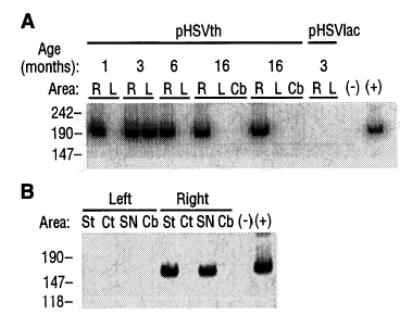
Persistence of pHSVth DMA and expression of human TH RNA. (A) DMA was extracted from sections and subjected to PCR with the use of primers specific to the human TH gene, and the products were electrophoresed (). Age is the time after gene transfer a rat was analyzed (6 months, rat pHSVth no. 27; 16 months, rats pHSVth no. 2, and no. 9). Brain areas: R, right injected striatum; L, left uninjected striatum; Cb, cerebellum. Minus sign indicates no DMA; plus sign indicates pHSVth DMA isolated from Escherichia coli, which should direct production of a 186-bp fragment (number of base pairs is shown at left). (B) RT-PCR analysis of RNA isolated from specific brain areas 1 month after injection of pHSVth () into the right striatum. Brain areas: St, striatum; Ct, cortex; SN, midbrain (substantia nigra); Cb, cerebellum. Minus sign indicates no RNA; plus sign indicates pHSVth DNA isolated from E. coli; the methods used () should generate a 160-bp fragment (number of base pairs is shown at left).
Comment in
-
Behavioral effects and gene delivery in a rat model of Parkinson's disease.Science. 1995 Aug 11;269(5225):856-7. doi: 10.1126/science.7638605. Science. 1995. PMID: 7638605 No abstract available.
References
-
- M. D. Yahr and K. J. Bergmann, Eds., Parkinson’s Disease (Raven, New York, 1987).
-
- Yahr MD, Duvoisin RC, Schear MJ, Barrett RE, Hoehn MM. Arch Neurol. 1969;21:343. - PubMed
Publication types
MeSH terms
Substances
Grants and funding
LinkOut - more resources
Full Text Sources
Other Literature Sources
Medical
