

Review
doi: 10.1002/jso.2930530503.
Hepatic regeneration and growth factors
Affiliations
- PMID: 7684910
- PMCID: PMC3005256
- DOI: 10.1002/jso.2930530503
Item in Clipboard
Review
Hepatic regeneration and growth factors
J Surg Oncol Suppl.
1993.
No abstract available
Figures
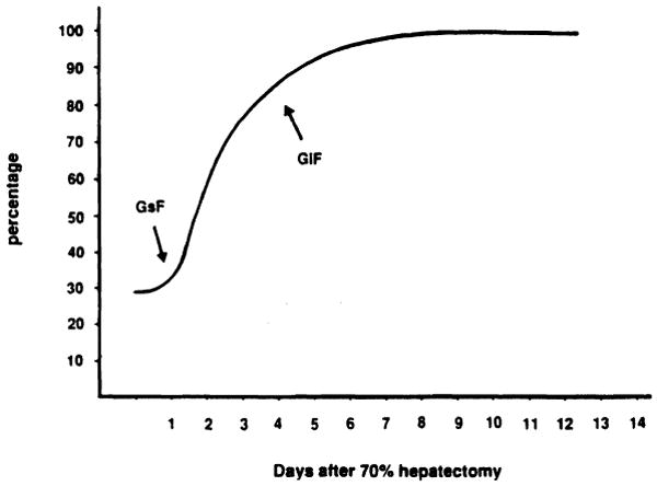
Liver mass recovery in rats after 70% hepatectomy. Liver resection was performed as described by Higgins and Anderson [15]. Liver mass is expressed as percentage of the liver weight in the intact animal. GsF, growth stimulatory factor; GiF, growth inhibitory factor.
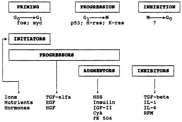
General pattern of the factors modulating liver regeneration and their time of intervention in the cell cycle. CyA, cyclosporin A; EGF, epidermal growth factor; HGF, hepatocyte growth factor; HSS, hepatic stimulatory substance; IGF, insulin-like growth factor; IL, interleukin; RPM, rapamycin; TGF, transforming growth factor.
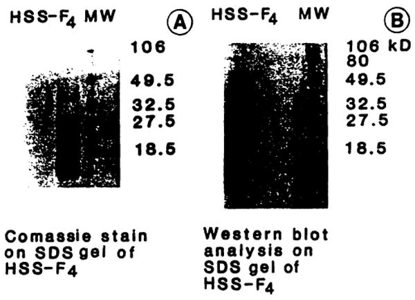
SDS-PAGE and Western blot analysis of acrylamide F4 (Ac-F4). For SDS-PAGE (A) final acrylamide concentration was 16%, in Tris 0.377 M, pH 8.8. Fifteen micrograms of protein underwent electrophoresis for 50 minutes at room temperature using 150 V, constant voltage, with Tris-HCI 0.125 M and 0.192 M glycine, pH 8.3, as reservoir buffer. Western blot analysis (B) was performed using an Immobilon membrane (Millipore). Protein transfer was conducted using 3-(cyclohexylamino)-1-propano-sulfonic acid (CAPS) 10 mM with 10% methanol, pH 11, as transfer buffer (90 minutes at 4°C with 70 V constant voltage). The membrane was blocked for 30 minutes with 3% gelatin in TTBS (Tris-buffered saline with 0.2% Tween 20) and incubated, for 60 minutes, with a specific monoclonal murine antibody against hepatic stimulatory substance (HSS) [8]. The membrane was then washed with TTBS and incubated with a second antibody in 1 % gelatin, against mouse immunoglobulins, carrying alkaline-phosphate substrate. The membrane was washed with TTBS and the immunoreactive band was identified.
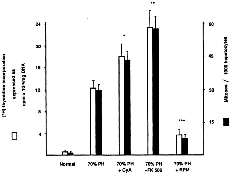
Effect of immunosuppressive agents on liver regeneration after 70% hepatectomy. [3H]thymidine incorporation and percentage of mitoses in normal and 70% hepatectomized rats treated or not treated with cyclosporin A (CyA), FK 506, and rapamycin (RPM). The animals were given oral CyA and intramuscular FK 506 or RPM at 10 mg, 1 mg, and 0.3 mg/kg body weight, respectively, for 3 days before surgery and again just after completing hepatic resection [15]. Values are the means from at least 15 rats ± SD. *, significantly different from 70% hepatectomized rats (P < .05). **, significantly different from 70% hepatectomized rats (P < .01) and CyA-treated 70% hepatectomized rats (P < .05). ***, significantly different from 70% hepatectomized rats (P < .01). Vehicle injections did not influence [3H]thymidine incorporation and percentage of mitoses in normal and 70% hepatectomized rats. PH, partial hepatectomy.
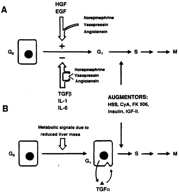
Possible mechanisms for the control of hepatic regeneration. The factors controlling hepatocyte proliferation are given, as described by Michalopoulos [1] (A) and Fausto [7] (B). Norepinephrine, vasopressin, and angiotensin potentiate GsF action and reduce GiF influence. For abbreviations, see Figure 2 legend.
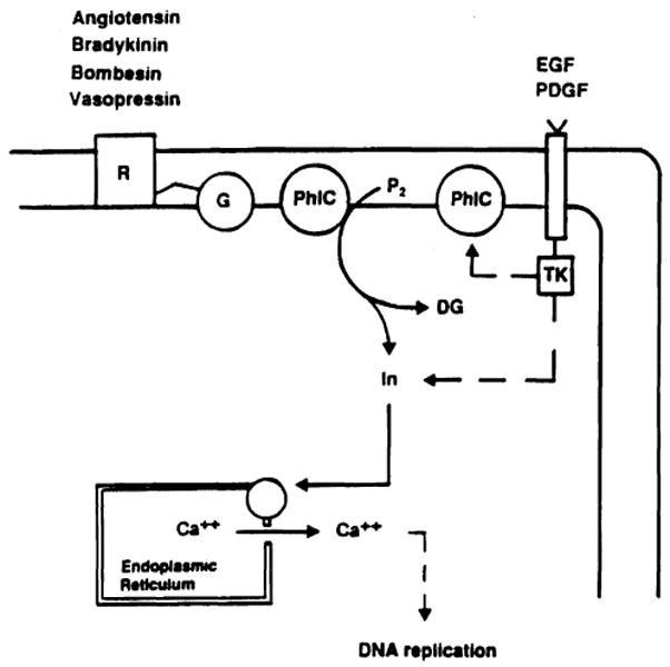
Signalling pathways of growth factors. R, cell surface receptor; G, guanine nucleotide binding protein; PhlC, phospholipase C; P2, phosphatidylinositol biphosphate; TK, tyrosine kinase; In, inositol; DG, diacylglycerol; PDGF, platelet-derived growth factor.
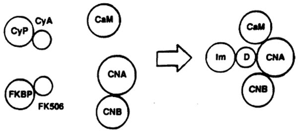
Interaction of CyA and FK 506 immunophilin complex with specific intracellular proteins. CyP, cyclophilin; FKBP, FK binding protein; CaM, calmodulin; CNA-CNB, calcineurin; Im, immunophilin; D, drug.
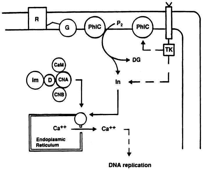
Intracellular signalling pathway of FK 506 and CyA. R, cell surface receptor; G, guanine nucleotide binding protein; PhlC, phospholipase C; P2, phosphatidylinositol biphosphate; TK, tyrosine kinase; In, inositol; DG, diacylglycerol; CaM, calmodulin; CNA-CNB, calcineurin; Im, immunophilin; D, drug.
References
-
- Michalopoulos G, Houck KA, Dolan ML, Luetteke NC. Control of hepatocvte replication by two serum factors. Cancer Res. 1984;44:4414–4419. - PubMed
-
- Goldberg M. Purification and partial characterization of a liver cell proliferation factor called hepatopoietin. J Cell Biochem. 1985;27:291–302. - PubMed
-
- Olsen PS, Boesby S, Kirkegaard P, et al. Influence of epidermal growth factor on liver regeneration after partial hepatectomy in rats. Hepatology. 1988;8:992–996. - PubMed
Publication types
MeSH terms
Substances
Grants and funding
LinkOut - more resources
Full Text Sources