Comparative Study
. 1993 Jul 1;151(1):473-88.

Human rheumatoid B-1a (CD5+ B) cells make somatically hypermutated high affinity IgM rheumatoid factors

Affiliations
Comparative Study

Human rheumatoid B-1a (CD5+ B) cells make somatically hypermutated high affinity IgM rheumatoid factors

L Mantovani et al. J Immunol. .

Abstract

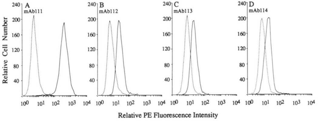
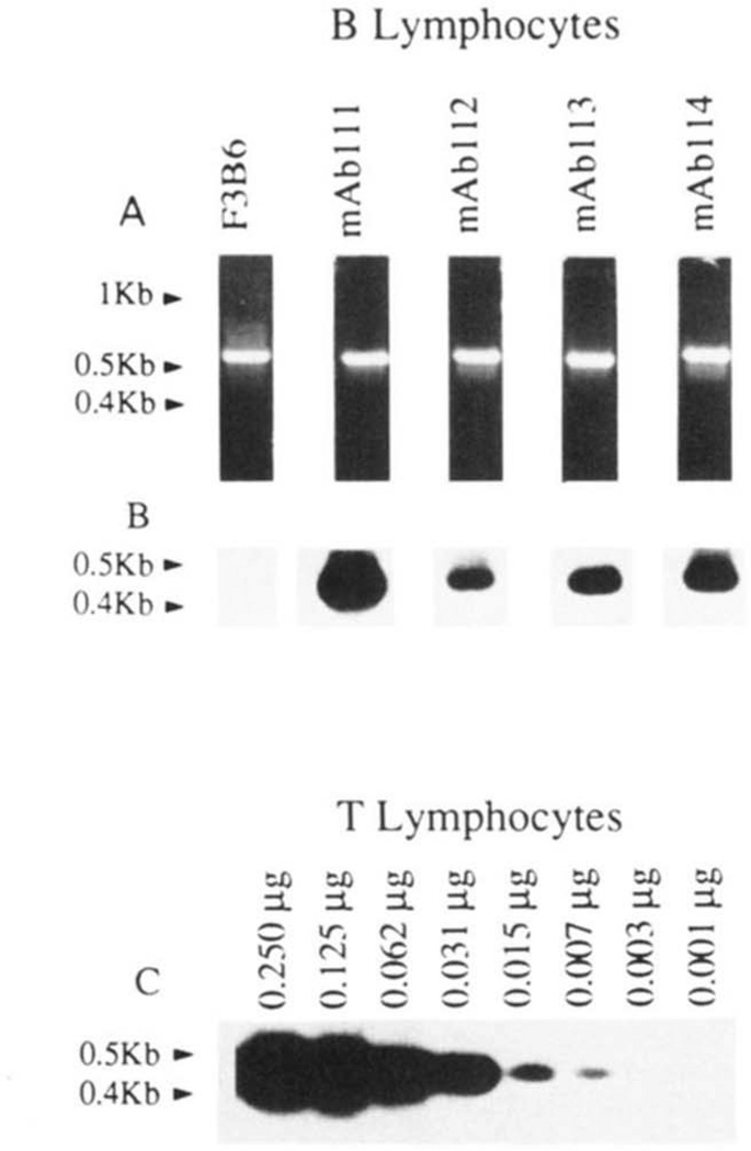
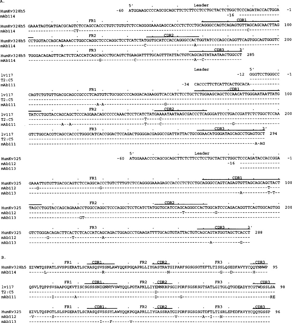
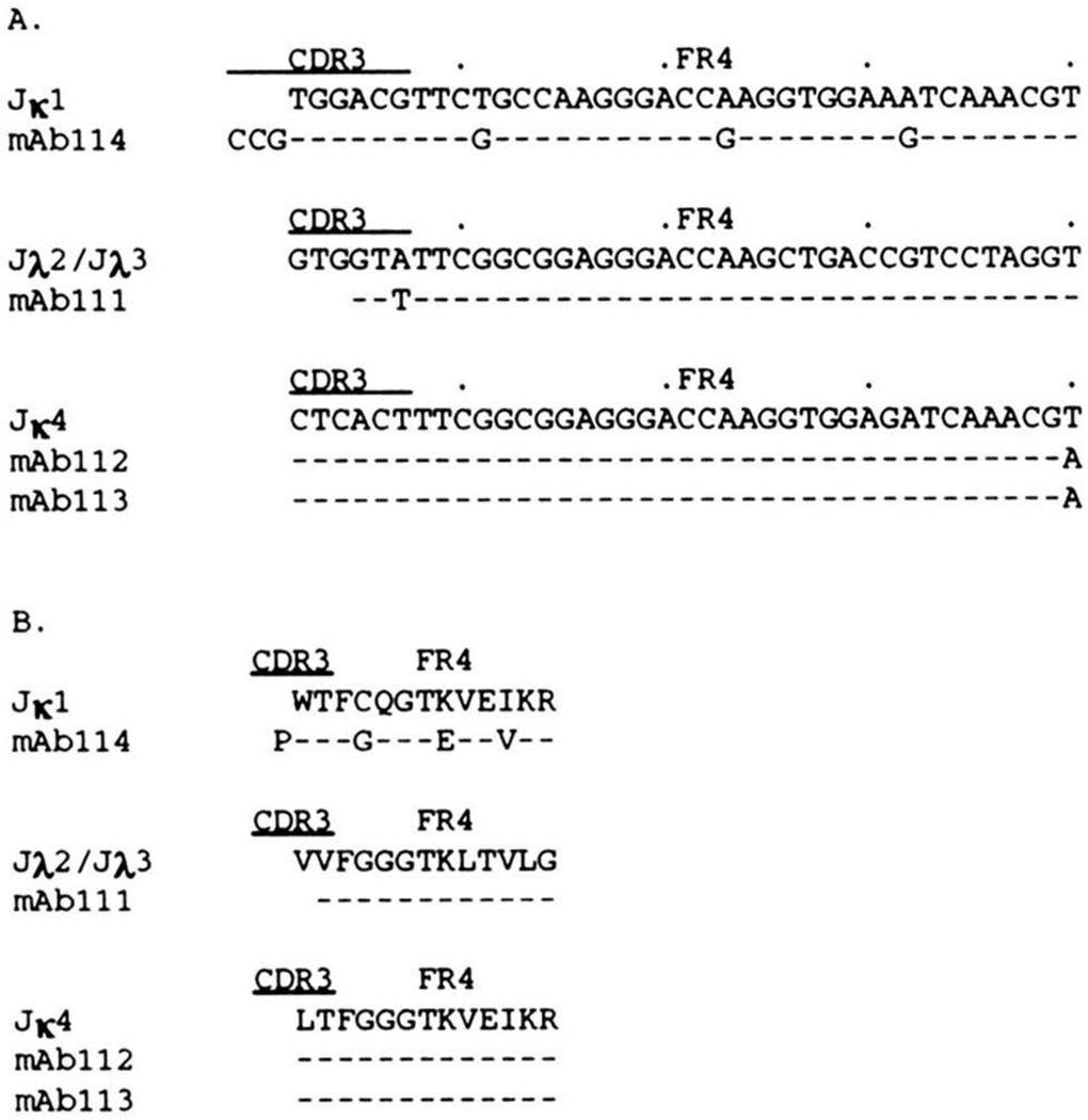
To analyze the structure and formally ascertain the B-1a cellular origin of IgM rheumatoid factor (RF) autoantibodies, we generated 4 IgM RF mAb-producing cell lines using sorted (surface CD5+) B-1a cells from a patient with active rheumatoid arthritis. The RF mAb111, mAb112, mAb113, and mAb114 were monoreactive and displayed a relatively high affinity for human IgG Fc fragment (Kd, 3.1 x 10(-7) to 6.8 x 10(-7) M). The B-1a origin of the lymphocytes that gave rise to the IgM RF was confirmed by the expression of surface CD5 and specific CD5 mRNA by all mAb-producing cell lines. Analysis of the genes encoding the RF mAb VH and VL regions revealed that members of the VHI and VHIII families were utilized in conjunction with V kappa IIIa, V kappa IIIb, or V lambda I genes. JH3 and JH4 genes were each utilized twice. The H chain CDR3 sequences were divergent and variable in length. The RF mAb VH genes were identical or closely related to those expressed in the "restricted" fetal B cell repertoire and/or were JH-proximal. For instance, mAb111 VH gene likely constituted a mutated variant of the expressed fetal 20P3 which is the second most JH-proximal gene (125 kb from JH). In addition, the expressed VH and VL genes were among those that have been found to encode other RF, different autoantibodies, high affinity antibodies induced by exogenous Ag, and natural autoantibodies in the adult and neonatal B cell repertoires. When compared with those of known germline genes, the expressed V gene sequences displayed a number of differences. By cloning and sequencing DNA from PMN of the same patient whose B lymphocytes were used for the mAb generation, we showed that such differences resulted from somatic hypermutation in the RF mAb112 VH gene. The germline gene (112GL) that presumably gave rise to the RF mAb112 VH segment was identical to the expressed fetal 51P1 gene. The distribution and the high replacement to silent mutation ratio of the nucleotide mutations in RF mAb112 VH segment were highly consistent with their selection by Ag. RF mAb113 was clonally related to RF mAb112, as shown by the utilization of the same sets of VHI-D-JH4 and V kappa IIIb-J kappa 4 genes, displaying identical junctional sequences, and the presence of two identical replacement and one silent mutations.(ABSTRACT TRUNCATED AT 400 WORDS)


Figures

FIGURE 1
FIGURE 1
Binding of the IgM RF mAb to solid-phase Ag, and Kd for IgC Fc fragment. The binding of each mAb to IgG Fc fragment, β-galactosidase from Escherichia coli, tetanus toxoid, ssDNA, human recombinant insulin, actin, and phosphorylcholine is expressed as optical absorbance at 492 nm.
FIGURE 2
FIGURE 2
Expression of surface CD5 by the RF mAb-producing B cells. EBV-transformed cells were reacted with biotinylated mouse IgG2a mAb to CD5 (solid lines) or with biotinylated mouse IgG2a mAb of irrelevant specificity (dotted lines), and then with PE-labeled streptavidin. Cells were analyzed by fluorescence flow cytometry.
FIGURE 3
FIGURE 3
Expression of CD5 mRNA by the RF mAb-producing B cells. mRNA from the EBV-transformed B cell lines (0.250 µg), T lymphocytes (0.250 to 0.001 µg), or the fusion partner F3B6 hybrid cells were reverse-transcribed. cDNA were amplified by PCR using β-actin or CD5-specific primers and fractionated on 1.2% agarose gel (see Materials and Methods). A, ethidium bromide-stained gel containing amplified β-actin DNA (~0.6 kb); B, hybridization of the 32P-labeled “internal” CD5-specific oligonucleotide probe with fractionated CD5 DNA amplified from cDNA reverse-transcribed from the mAb-producing cell lines or the fusion partner F3B6 cells; C, hybridization of 32P-labeled CD5-specific oligonucleotide probe to fractionated DNA amplified from cDNA independently reverse-transcribed from different amounts (0.250 to 0.001 µg) of purified T cell mRNA.
FIGURE 4
FIGURE 4
Nucleotide (A) and deduced amino acid (B) sequences of the VH genes utilized by the IgM RF mAb. In each cluster, the top sequence is given for comparison and represents the germline VH gene displaying the highest identity to the expressed VH genes. The HHG19 VH gene belongs to the VHIII family. VI-2 and hv1263 are members of the VHI gene family. Dashes indicate identities. Solid lines above each cluster encompass CDR sequences. Small letters (in the case of hv1263 and 112GL) denote intron sequences. 112GL is the germline sequence we amplified from PMN DNA of the patient whose B cells were used for the generation of mAb112. Sequences encompassed by the oligonucleotide primers are underlined. The present sequences are available from EMBL/GenBank/DDBJ under accession numbers L14453, L14454, L14455, and L14456.
FIGURE 5
FIGURE 5
Nucleotide (A) and deduced amino acid (B) sequences of the D and JH segments of the RF mAb. Germline D genes are given for comparison. Dashes indicate identities. Inverted DN4 sequence is the reverse complement of the germline DN4 gene sequence. The deduced amino acid sequences are divided in CDR3 and FR4. The present sequences are available from EMBL/GenBank/DDBJ under accession numbers L14453, L14454, L14455, and L14456.
FIGURE 6
FIGURE 6
Nucleotide (A) and deduced amino acid (B) sequences of the VL genes utilized by IgM RF mAb. The top sequence in each cluster is used for comparison. Identities are indicated by dashes. Solid lines above each cluster encompass CDR sequences. Sequences encompassed by the oligonucleotide primers are underlined. The humKv328h5 and humKv325 genes belong to the VκIIIa and VκIIIb subgroups, respectively. The Iv117 and T2:C5 genes belong to the VλI subgroup. The present sequences are available from EMBL/GenBank/DDBJ under accession numbers L14451, L14452, L14457, and L14458.
FIGURE 7
FIGURE 7
Nucleotide (A) and deduced amino acid (B) sequences of the JL segments used by RF mAb. The top germline sequence in each cluster is used for comparison. Identities are indicated by dashes. Solid lines above each cluster encompass CDR3 sequences. The present sequences are available from EMBL/GenBank/DDBJ under accession numbers L14451, L14452, L14457, and L14458.
FIGURE 8
FIGURE 8
Evidence for somatic mutations in the mAb112 VH gene. A, ethidium bromide staining of amplified DNA after fractionation in agarose gel electrophoresis. Using the CDR1 (112CDR1) and the FR3 (HI-7) oligonucleotide primers (see Materials and Methods), a PCR amplification product of appropriate size was obtained by priming DNA from the mAb112-producing cell line (hybridoma DNA) but not from autologous PMN (PMN DNA). B, ethidium bromide staining of amplified DNA after fractionation in agarose gel electrophoresis. Using the leader (HI-6) and FR3 (HI-7) sequence oligonucleotide primers, amplification products of identical size were obtained by priming DNA from both mAb112 hybridoma (hybridoma DNA) and autologous PMN (PMN DNA). C, Southern blot hybridization of the PCR products shown in B with the 32P-labeled oligonucleotide 112CDR1 probe encompassing the entire CDR1 of the mAb112 VH gene. A strong, positive hybridization signal was detected only with DNA amplified from the mAb112-producing B cell line.

Similar articles

Cited by

References

    1. Carson DA, Chen PP, Fox RI, Kipps TJ, Jirik F, Goldfien RD, Silverman G, Radoux V, Fong S. Rheumatoid factors and immune networks. Annu. Rev. Immunol. 1987;5:109. - PubMed
    1. Chen PP, Silverman GP, Liu M-F, Carson DA. Idiotypic and molecular characterization of human rheumatoid factors. In: Carson DA, Chen PP, Kipps TJ, editors. Idiotypes in Biology and Medicine. Vol. 48. Basel: Chem. Immunol., S. Karger; 1990. p. 63. - PubMed
    1. Chen PP, Olsen NJ, Yang P-M, Soto-Gil RW, Olee T, Siminovitch KA, Carson DA. From human autoantibodies to fetal antibody repertoire to B cell malignancy: it’s a small world after all. Int. Rev. Immunol. 1990;5:239. - PubMed
    1. Avrameas S. Natural autoantibodies: from “horror autotoxicus” to “gnothi seauton.”. Immunol. Today. 1991;12:154. - PubMed
    1. Riboldi P, Kasaian MT, Mantovani L, Ikematsu H, Casali P. Natural antibodies. In: Bona CA, Siminovitch K, Zanetti M, Theofilopoulos AN, editors. The Molecular Pathology of Autoimmune Diseases. Philadelphia, PA: Gordon and Breach Science Publishers; 1993. pp. 45–64.

Publication types

MeSH terms

LinkOut - more resources