. 1993 Nov 4;366(6450):69-72.
doi: 10.1038/366069a0.

Spontaneous loss of T-cell tolerance to glutamic acid decarboxylase in murine insulin-dependent diabetes

Affiliations

Spontaneous loss of T-cell tolerance to glutamic acid decarboxylase in murine insulin-dependent diabetes

D L Kaufman et al. Nature. .

Abstract

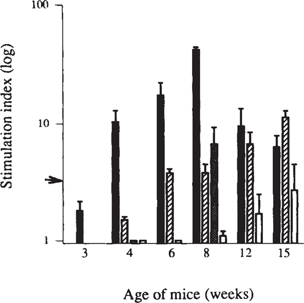
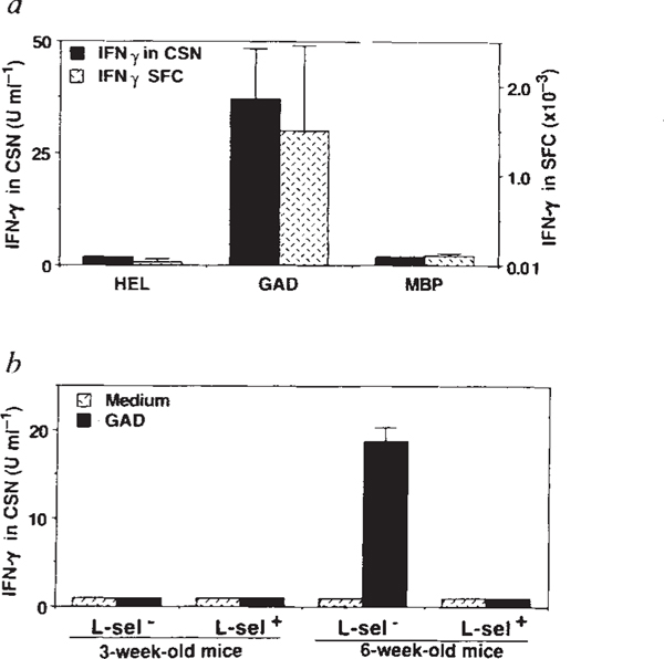
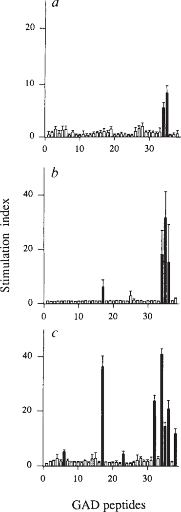
Insulin-dependent diabetes mellitus (IDDM) in non-obese diabetic (NOD) mice results from the T-lymphocyte-mediated destruction of the insulin-producing pancreatic beta-cells and serves as a model for human IDDM. Whereas a number of autoantibodies are associated with IDDM, it is unclear when and to what beta-cell antigens pathogenic T cells become activated during the disease process. We report here that a T-helper-1 (Th1) response to glutamate decarboxylase develops in NOD mice at the same time as the onset of insulitis. This response is initially limited to a confined region of glutamate decarboxylase, but later spreads intramolecularly to additional determinants. Subsequently, T-cell reactivity arises to other beta-cell antigens, consistent with intermolecular diversification of the response. Prevention of the spontaneous anti-glutamate decarboxylase response, by tolerization of glutamate decarboxylase-reactive T cells, blocks the development of T-cell autoimmunity to other beta-cell antigens, as well as insulitis and diabetes. Our data suggest that (1) glutamate decarboxylase is a key target antigen in the induction of murine IDDM; (2) autoimmunity to glutamate decarboxylase triggers T-cell responses to other beta-cell antigens, and (3) spontaneous autoimmune disease can be prevented by tolerization to the initiating target antigen.


Figures

FIG. 1
FIG. 1
Proliferative T-cell responses to β-cell antigens develop spontaneously in NOD mice in a defined chronological order. Antigen-induced blastogenesis was measured in spleen cells from newborn to 28-week-old female NOD mice (data from 3–15 weeks are shown). β-Cell antigens include GAD65 (black bars), Hsp65 peptide PALDSLTPANED (striped bars), carboxypeptidase H (grey bar) and insulin (white bars). Data are expressed as stimulation indices (SI) ± standard error of the mean (s.e.m.), calculated from 3–5 mice tested individually in 2 separate experiments for each time point. Arrow indicates SI = 3, the level of significance. Carboxypeptidase H responses were only tested at 4 and 8 weeks. None of the control antigens (hen egg-white lysozyme, human serum albumin, E. coli β-galactosidase or murine myelin basic protein) induced T-cell proliferation at any age. Also, none of the β-cell antigens or control antigens induced proliferation of T cells from age-matched control BALB/c or (NOD × BALB/c) F1 mice (data not shown). METHODS. NOD (Taconic farms) and BALB/c mice (Jackson Laboratories) were housed under specific pathogen-free conditions. Spleen cells were tested directly ex vivo for proliferative recall responses. Cells were plated at 1 × 10⁶ cells per well in 96-well microtitre plates in 200 μl HL-1 medium (Ventrex) containing 2 mM glutamine and 10 μg ml−1 antigen, or 7 μM peptide (the optimal concentrations for all time points tested) in triplicate. During the last 16 h of the 72 h culture period, 1 μCi [3H]thymidine was added per well. Incorporation of label was measured by liquid scintillation counting. Human GAD65 (ref. 25) and E. coli β-galactosidase were both purified from recombinant bacteria using a hexahistidine tag and metal-affinity chromatography. Bovine carboxypeptidase H was a generous gift from L. Fricker; human insulin was purchased from Eli Lilly.
FIG. 2
FIG. 2
GAD-specific T cells in 6–9-week-old female NOD mice are primed Thl type CD4+ lymphocytes based on their production of IFN-γ (a and b), enhanced clonal size (a) and L-selectin phenotype (b). a, Detection of IFN-γ by ELISA in culture supernatants (CSN) of spleen cells from 6–9-week-old mice 48 h after challenge with GAD or control antigens hen egg-white lysozyme (HEL) and murine myelin basic protein (MBP). High concentrations of IFN-γ were detected only in cultures containing GAD. IFN-γ production was measured by ELISA using IFN-γ-specific monoclonal antibodies R4–6A2 and XMG 1.2 (Pharmingen). T cells from age-matched BALB/c mice did not respond to GAD or to control antigens (data not shown). The frequency of antigen-specific IFN-γ-producing cells was determined by an ELISA spot technique using the complementary IFN-γ mAbs. Frequency of antigen-induced spot-forming cells (SFC) is shown. Values are the mean ±s.e.m. from 5 female NOD mice, tested in triplicate cultures, with and without antigen. Results are from a single experiment and are representative of 3 experiments. b, Detection of GAD-specific, IFN-γ-producing cells in the L-selectin (L-sel) subpopulation of CD4+ cells. CD4+ cells were isolated from the spleens by panning on goat-anti-mouse immunoglobulin (Zymed) and on anti-CD8 (mAb 58.6–72)-coated plates. CD4+ cells were subfractionated using L-selectin-specific mAb MEL-14. CD4+ L-selectin+ and CD4+ L-selectin fractions were seeded in triplicate at 2 × 10⁵ cells per well with irradiated (3,000 rad) spleen cells of 3-week-old NOD mice (5 × 10⁵ cells per well) as a source of antigen-presenting cells. GAD-induced IFN-γ production was measured after 48 h by ELISA.
FIG. 3
FIG. 3
Intramolecular spreading of autoimmunity within the GAD molecule. Spleen cells were tested from 4-week-old (a), 5-week-old (b) and 7-week-old (c) NOD mice for proliferative responses to GAD65 peptides. A set of 38 peptides, each 20–23 amino acids long, span the entire human GAD65 (ref. 25) molecule with overlaps of 5 amino acids. These peptides are numbered successively from the N terminus. Peptides that triggered stimulation indices >3 are indicated as black bars. These peptides did not induce proliferation in T cells from NOD mice <3 or >16 weeks in age (paralleling reactivity to whole GAD), or from control (BALB/c × NOD)F1 mice (data not shown). Data are represented as the mean SI ±s.e.m. calculated from 3–6 mice tested individually in two separate experiments for each age group. METHODS. Proliferation was assayed as described for Fig. 1. Peptides were present in cultures at 7 μM and label was added during the last 16 h of a 5-day culture. Peptides were synthesized using standard F-moc chemistry and purified by reverse-phase HPLC (Advanced Chem-tech). The sequences of stimulatory peptides are: peptide 509–528 (no. 34), (IPPSLRYLED–NEERMSRLSK); peptide 524–543 (no. 35), (SRLS-KVAPVIKARMMEYGTT); and peptide 247–266 (no. 17), (NMYAMMIARFKMFPEVKEKG). Murine and human GAD65 share 95% amino-acid identity and are 98% conserved. Underlined amino acids are conservatively substituted in murine GAD65. In separate experiments, the murine forms of these peptides were tested and produced similar results.

Comment in

References

    1. Castaño L. & Eisenbarth GS A. Rev. Immun 8, 647–679 (1990). - PubMed
    1. Palmer JP Diabetes, Rev. 1,104–115 (1993).
    1. Kaufman DL, McGinnis JF, Krieger NR & Tobin AJ Science 232, 1138–1140 (1986). - PMC - PubMed
    1. Erlander MG, Tillakaratne NJK, Feldblum S, Patel N. & Tobin AJ Neuron 7, 91–100 (1991). - PubMed
    1. Kaufman DL & Tobin AJ Trends pharmac. Sei 14, 107–109 (1993). - PubMed

Publication types