. 1995 Apr;21(4):1070-8.

Cell proliferation and oncogene expression after bile duct ligation in the rat: evidence of a specific growth effect on bile duct cells

Affiliations

Cell proliferation and oncogene expression after bile duct ligation in the rat: evidence of a specific growth effect on bile duct cells

L Polimeno et al. Hepatology. 1995 Apr.

Abstract

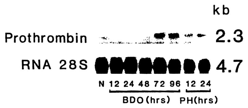
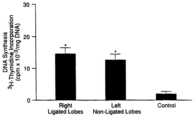
The proliferative response of the rat liver was measured after temporary or permanent total biliary obstruction (BDO) and in different regions after selective ligation of the lobar ducts draining the right 60% of the hepatic mass. The results were compared with those after 70% partial hepatectomy (PH). Cell proliferation was assessed globally by measuring DNA synthesis and stratified to the separate cell populations with cytostaining techniques that allowed distinction of hepatocytes, duct cells, and nonparenchymal cells (NPCs). In selected experimental groups, gene expression was determined of transforming growth factor-beta 1 (TGF beta-1), prothrombin, c-erb-B2, transforming growth factor alpha (TGF alpha), human Cyclophilin (CyP), and 28S ribosomal RNA. The stimulation of a proliferative response to total BDO required obstruction for longer than 24 hours, but after this deligation did not switch off regeneration. In the first week after permanent BDO, there was progressive infiltration of NPCs, fibrous linkage of some portal areas, and a crescendo of DNA synthesis that was obvious at 24 hours, maximal at 48 hours, and back nearly to baseline at 6 days. At the 2-day mark, the bile duct cells had a 17-fold increase in proliferation, accompanied by a threefold to fourfold increase in hepatocyte renewal. Little or no increase in expression of TGF alpha or the hepatocyte-specific prothrombin gene was detectable in the first 48 hours, whereas levels of the oncogene c-erb-B2 that is associated with cholangiocarcinoma were expressed from 48 to 96 hours.(ABSTRACT TRUNCATED AT 250 WORDS)


Figures

Fig. 1
Fig. 1
Regional biliary duct obstruction procedure that permits the study of obstructed and non-obstructed liver in the rat. The drained non-cholestatic portion is approximately 40% of the liver mass.
Fig. 2
Fig. 2
Evidence of lack of ductal cross-communication between obstructed and non-obstructed lobes proved with methylene-blue injection into the common bile duct. Right lobe obstructed and left lobe nonobstructed.
Fig. 3
Fig. 3
Rat liver 7 days after common bile duct ligation. Proliferation of bile ducts and nonparenchimal liver cells, that form bridging connection between portal areas. (Trichrome stain; original magnification × 100)
Fig. 4
Fig. 4
DNA synthesis (mean ± SD) on successive days after total bile duct obstruction (n = 5 rats at each time point). *p < .005 VS. 0 time control.
Fig. 5
Fig. 5
Rat liver 6 days after common bile duct ligation. BrdU incorporation in proliferating bile duct epithelial cells (circled) and hepatocytes. (Immunostain with BrdU-antibodies and hematoxylin; original magnification ×400.)
Fig. 6
Fig. 6
Percent (mean ± SD) of hepatocytes and bile duct cells with nuclear labeling with BrdU at successive times after total bile duct obstruction (upper) and 70% hepatectomy (lower) (n = 3 rats each time point). The control value (0 time) is the mean average of five sham-operated rats. *P < .005 vs. sham controls. †P < .005 vs. value of PH rats at same time point.
Fig. 7
Fig. 7
Effect of total biliary obstructions for different durations on the percent of BrdU nuclear labeling of duct cells (mean ± SD) in animals killed at 2 days (n = 5 rats at each time point). *P < .005 vs. value at time 0.
Fig. 8
Fig. 8
Northern blot analysis of RNA from rat liver at different times after PH and BDO, and hybridized to probes for TGFα or cyclophilin (n = 3 rats for each time point).
Fig. 9
Fig. 9
Northern blot analysis of RNA from rat liver at different times after PH or BDO, and hybridized to probes for c-raf or c-erb-B2.
Fig. 10
Fig. 10
Northern blot analysis of RNA from rat liver at different times after PH and BDO, and hybridized to the probe of prothrombin.
Fig. 11
Fig. 11
Northern blot analysis of RNA from rat liver at different times after PH and BDO, and hybridized to the probe of TGF-β1.
Fig. 12
Fig. 12
DNA synthesis (mean ± SD) in ligated and nonligated liver lobes of rats with regional biliary tree obstruction (n = 10 rats for the experimental bars and 5 for controls). *P < .005 vs. controls.
Fig. 13
Fig. 13
Ligated lobe of rat liver 48 hours after regional bile duct ligation. BrdU incorporation in proliferating bile duct epithelial cells and hepatocytes. *P < .05 vs. hepatocytes in sham operated rats. **P < .01 VS. hepatocytes in nonligated lobes. (lmmunostain with BrdU-antibodies and hematoxylin; original magnification ×400.)

References

    1. Tramas EG, Symeonidis A. Morphologic and functional changes in the livers of rats after ligation or excision of the common bile duct. Am J Pathol. 1957;33:13–27. - PMC - PubMed
    1. Accatino L, Contreras A, Fernandez S, Quintana C. The effect of complete biliary obstruction on bile flow and bile acid excretion: postcholestatic choleresis in the rat. J Lab Clin Med. 1979;93:706–717. - PubMed
    1. Alpini G, Lenzi R, Sarkozi L, Tavoloni N. Biliary physiology in rats with bile ductular cell hyperplasia: evidence for a secretory function of proliferated bile ductules. J Clin Invest. 1988;81:569–578. - PMC - PubMed
    1. Slott PA, Liu MH, Tavoloni N. Origin, pattern and mechanism of bile duct proliferation following biliary obstruction in the rat. Gastroenterology. 1990;99:466–477. - PubMed
    1. Shibayama Y. Factors producing bile infarction and bile duct proliferation in biliary obstruction. J Pathol. 1990;160:57–62. - PubMed

Publication types