. 1995 Feb 15;87(4):280-5.
doi: 10.1093/jnci/87.4.280.

Molecular characterization of defective antigen processing in human prostate cancer

Affiliations

Molecular characterization of defective antigen processing in human prostate cancer

M G Sanda et al. J Natl Cancer Inst. .

Abstract

Background: Gene-modified tumor cell vaccines have shown efficacy in animal models of malignancy, including prostate cancer. Class I major histocompatibility complex (MHC) assembly and function in the cellular targets of such therapies is pivotal in determining the efficacy of specific cytokine-secreting tumor vaccines.

Purpose: To help guide development of genetically engineered vaccine therapy for human prostate cancer, potential immune resistance pathways were evaluated by analysis of class I MHC assembly in prostate cancer cells.

Method: Class I MHC assembly in metastasis-derived human prostate cancer cell lines (LNCaP, PPC-1, DU-145, PC-3, and TSU) and a normal prostate-derived cell line (TP-2) were characterized by phenotypic, molecular, and functional assays. Assembled class I MHC and antigen was measured by flow cytometry; mRNA levels of assembly components (class I MHC heavy chain, beta 2-microglobulin, and the antigen transporter gene product TAP-2) were determined; and antigen processing was measured with a chimeric reconstituted system using vaccinia vectors. Restoration of antigen processing was attempted by interferon gamma stimulation and by transfection with mouse class I MHC heavy-chain cDNA.

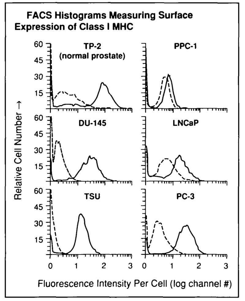
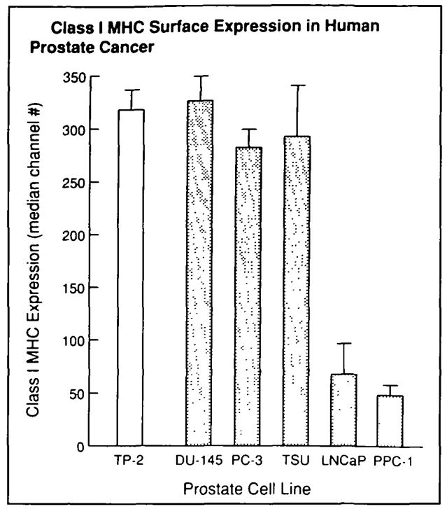
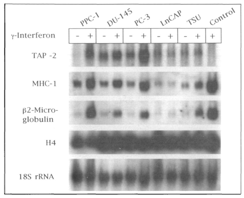
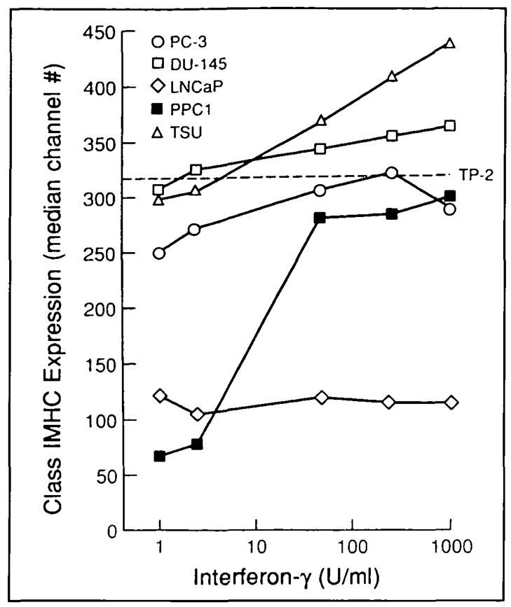
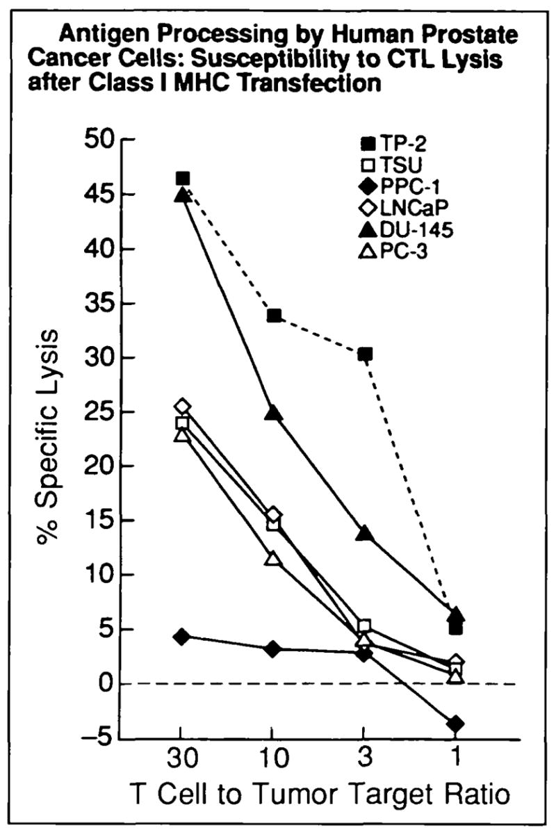
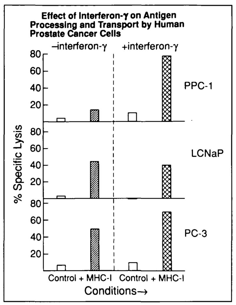
Results: Assembled class I MHC was underexpressed in two (LNCaP and PPC-1) of five prostate cancer cell lines compared with normal prostate-derived controls. PPC-1 cells underexpressed TAP-2 mRNA despite abundant class I MHC and beta 2-microglobulin message. Induction of TAP-2 by interferon gamma indicated that coding sequences for TAP-2 message were present in PPC-1. Resistance to cytotoxic T lymphocytes (CTL) lysis showed a functional defect in antigen transport by PPC-1 cells; reversal of the molecular defect with interferon gamma led to restoration of functional antigen processing. In contrast, LNCaP cells had competent antigen transport but deficient class I MHC heavy-chain function despite abundant class I MHC RNA; though refractory to stimulation by interferon gamma, this defect responded to transfection of class I MHC heavy-chain cDNA.

Conclusions: Metastatic prostate cancer cells can escape T-cell recognition via divergent mechanisms of defective class I MHC assembly. The specific underexpression of TAP-2 gene product in PPC-1 cells contrasts with prior studies of TAP gene underexpression in lung cancer (which concurrently underexpressed class I MHC heavy chain) and provides evidence for a regulatory pathway controlling TAP-2 gene expression in human cancers that may not affect class I MHC heavy-chain expression.

Implications: In clinical application of gene therapy for prostate cancer, these findings provide a rationale for focusing on strategies that can circumvent sole reliance on class I MHC-mediated tumor cell recognition by CTL.


Figures

Fig. 1
Fig. 1
Underexpression of cell-surface class I MHC by human prostate cancer cells. FACS analysis was used to measure class I MHC expression in five cultured human prostate cancer cell lines (DU-145, LNCaP, PC-3, PPC-1, and TSU) and one immortalized cell line derived from normal prostate epithelium (TP-2) as described in the “Materials and Methods” section. Representative FACS histograms: solid lines indicate staining with human class I MHC-specific antibody (w6/32), dashed lines indicate control histograms with fluorescein-conjugated, isotype-matched control antibody determining background fluorescence. Three separate experiments were performed with similar results.
Fig. 2
Fig. 2
Relative expression of class I MHC was determined by subtracting median fluorescence of isotype-matched controls from median fluorescence of class I MHC antibody-labeled cells. Mean values from three experiments are shown for each cell line; error bars represent SE. LNCaP and PPC-1 were found to have diminished expression of class I MHC relative to the normal prostate control line TP-2. (Paired t test with Bonferroni adjustment for multiple comparisons: LNCaP, P<.025; PPC-1, P<.005).
Fig. 3
Fig. 3
Northern blot hybridization of antigen processing components in human prostate cancer cell lines. RNA from five human prostate cancer cell lines (PPC-1, DU-145, PC-3, LNCaP, and TSU; with and without stimulation by interferon gamma [γ-interferon]) and a control somatic cell-fusion product cell line with deletion of the TAP-2 gene (T-2 control stimulated by interferon gamma) was purified, transferred to nylon membrane, and hybridized with 32P-labeled probes for TAP-2, class I MHC, β2-microglobulin, and, to control for uniformity in RNA loading and transfer, histone H4 mRNA and 18S rRNA. Autoradiographs of each hybridization are shown.
Fig. 4
Fig. 4
Defective class I MHC expression in human prostate cancer restored using interferon gamma (interferon-γ). Surface expression of class I MHC was measured by FACS 24 hours after culture of human prostate cancer cell lines with interferon gamma. Base-line class I MHC expression by normal prostate-derived TP-2 (dashed line) is shown as a reference standard. PPC-l cells demonstrate a significant, dose-dependent response to interferon gamma (P<.01; t test with pooled variance), whereas LNCaP underexpression of class I MHC is refractory to interferon gamma. Responses to interferon gamma appeared after 8 hours of culture, and stimulation for up to 3 days revealed similar responses (not shown). Three experiments were performed with similar results.
Fig. 5
Fig. 5
Class I MHC-mediated antigen processing by human prostate cancer. Class I MHC-restncted T-cell killing targeted via a model viral antigen was used t o measure antigen processing and transport by normal and cancerous human prostate. Class I MHC-restricted, vaccinia antigen-specific lysis (y axis vaccinia antigen presented by mouse Kd class I MHC transfected into tumor target cells using vaccinia vector) was measured by chromium release. Dashed line indicates normal prostate control cells; solid lines indicate metastasis-derived human prostate cancer cells. Each data point represents a mean of triplicate measurements (SE consistently <10%); this experiment was repeated twice with similar results. T-cell killing of all lines except PPC-1 was significant (P<.01).
Fig. 6
Fig. 6
Prostate cancer cell lines PPC-1, LNCaP, and PC-3 were cultured with or without 250 U/mL of interferon gamma (interferon-γ) for 24 hours, and their susceptibility to CTL-mediated killing was then measured. Parallel conditions are identified on the x axis: clear columns (control lysis) = lysis of cells transfected with wild-type vaccinia (lacking Kd) or nonspecific, background lysis (with or without interferon gamma stimulation of targets prior to transfection); single-hatched columns (class I MHC-specific lysis) = lysis of cancer cells transfected with vac-Kd; cross-hatched columns (class I MHC-specific lysis) = lysis of cancer cells transfected with vac-Kd after interferon gamma treatment. Tumor lysis (T cell to tumor ratio = 30.1) is shown on y axis for each condition; each value represents a mean of triplicate measurements (SE consistently <10%). Lysis was T-cell dependent as in Fig. 5 (10:1, 3:1, and 1:1 dilutions not shown here). Increase in PPC-1 killing after interferon gamma treatment was statistically l significant (P<.005; pooled variance t test).

Comment in

References

    1. Walsh PC. Prostate cancer kills: a strategy to reduce deaths. Urology. 1994;44:463–466. - PubMed
    1. Golumbek PT, Lazenby AJ, Levitsky HI, et al. Treatment of established renal cell cancer by tumor cells engineered to secrete interleukin-4. Science. 1991;254:713–716. - PubMed
    1. Asher AL, Mulé JJ, Kasid A, et al. Murine tumor cells transduced with the gene from tumor necrosis factor-alpha. Evidence for paracrine immune effects of tumor necrosis factor against tumors. J Immunol. 1991;146:3227–3234. - PMC - PubMed
    1. Restifo NP, Spiess PJ, Karp SE, et al. A nonimmunogenic sarcoma transduced with the cDNA for interferon gamma elicits CD8+ T cells against the wild-type tumor: correlation with antigen presentation capability. J Exp Med. 1992;175:1423–1431. - PMC - PubMed
    1. Trojan J, Johnson TR, Rudin SD, et al. Treatment and prevention of rat glioblastoma by immunogenic C6 cells expressing antisense insulin-like growth factor I RNA. Science. 1993;259:94–97. - PubMed

Publication types

Substances