. 1994 Nov;120(11):3131-43.
doi: 10.1242/dev.120.11.3131.

Transcriptional regulation of string (cdc25): a link between developmental programming and the cell cycle

Affiliations

Transcriptional regulation of string (cdc25): a link between developmental programming and the cell cycle

B A Edgar et al. Development. 1994 Nov.

Abstract

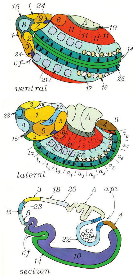
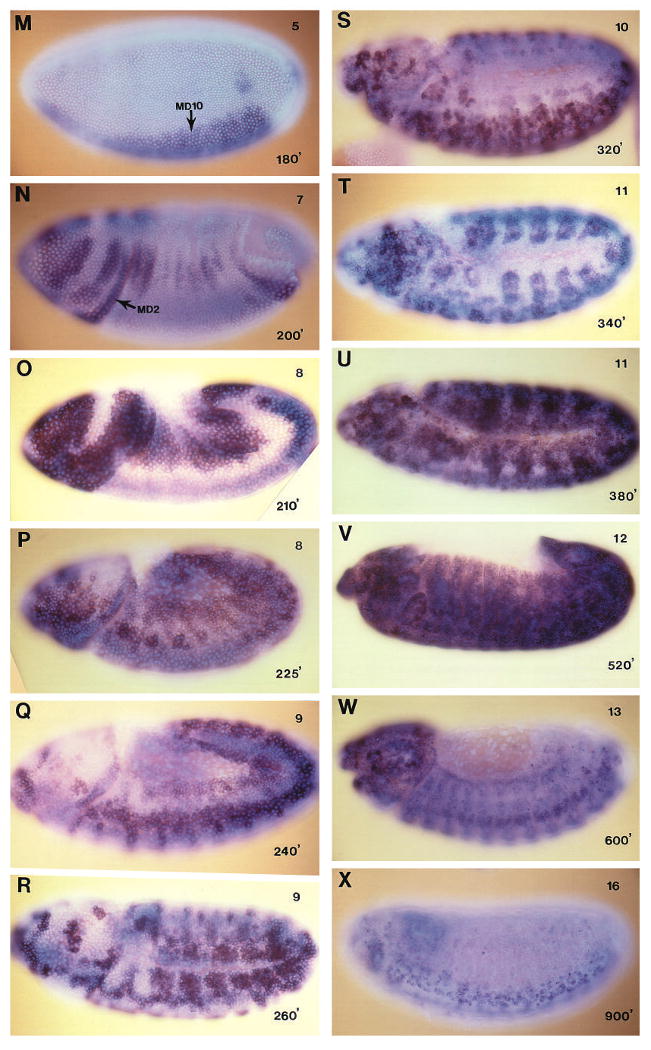
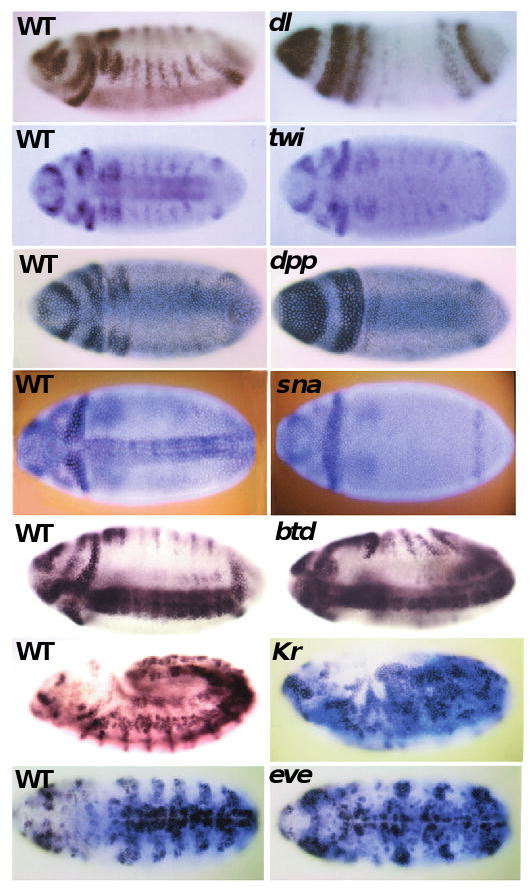
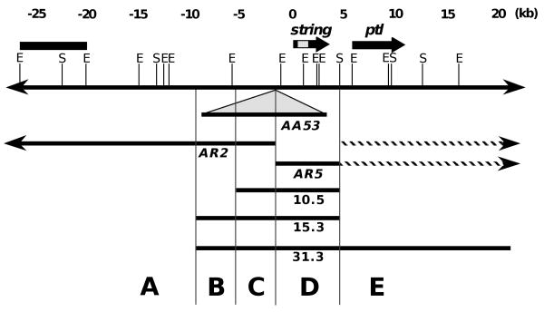
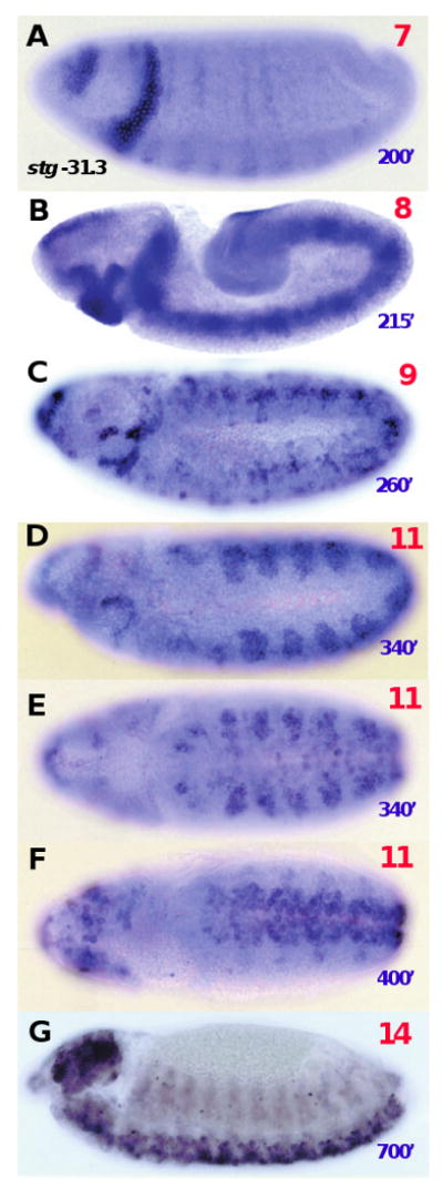
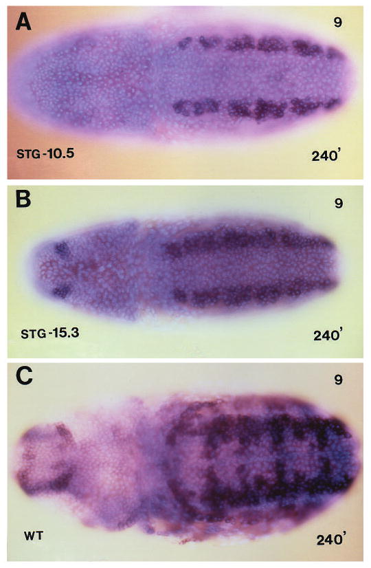
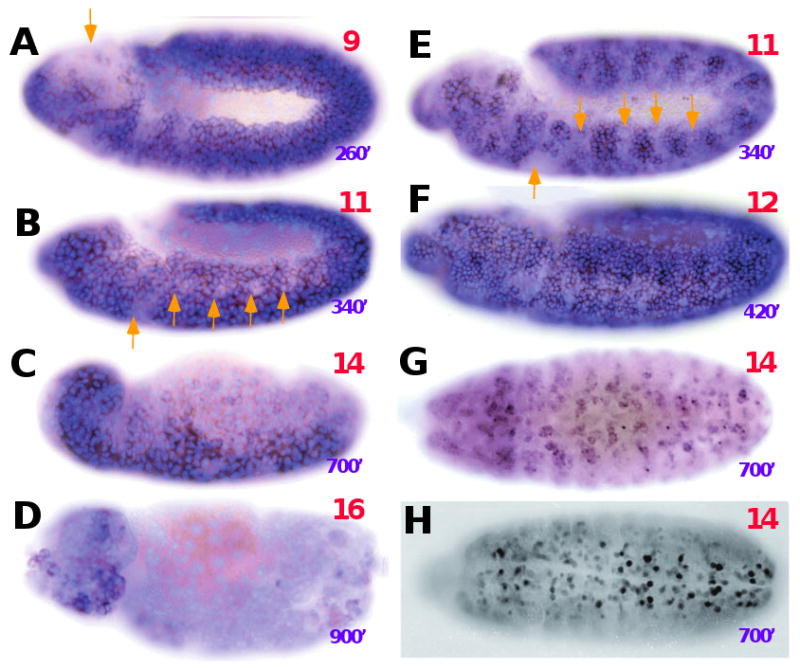
During postblastoderm embryogenesis in Drosophila, cell cycles progress in an invariant spatiotemporal pattern. Most of these cycles are differentially timed by bursts of transcription of string (cdc25), a gene encoding a phosphatase that triggers mitosis by activating the Cdc2 kinase. An analysis of string expression in 36 pattern-formation mutants shows that known patterning genes act locally to influence string transcription. Embryonic expression of string gene fragments shows that the complete pattern of string transcription requires extensive cis-acting regulatory sequences (> 15.3 kb), but that smaller segments of this regulatory region can drive proper temporal expression in defined spatial domains. We infer that string upstream sequences integrate many local signals to direct string's transcriptional program. Finally, we show that the spatiotemporal progression of string transcription is largely unaffected in mutant embryos specifically arrested in G2 of cycles 14, 15, or 16, or G1 of cycle 17. Thus, there is a regulatory hierarchy in which developmental inputs, not cell cycle inputs, control the timing of string transcription and hence cell cycle progression.


Figures

Fig. 1
Fig. 1
Map of the mitotic domains of cycle 14. Mitotic domains are indicated in distinct colors from ventral (top) lateral (middle) and sagittal section (bottom) perspectives. Each mitotic domain represents a group of cells that divide with similar timing. The domains are numbered according to the order in which mitosis occurs during cycle 14. However, some of the earliest cycle 15 divisions occur before the last cycle 14 divisions. The lateral view also includes segmental indicators along the ventral (lower) surface and the expression of engrailed (stippled), which occurs in the posterior region of each segment. This map was reproduced from Foe (1989), who mapped the division patterns using distributions of mitotic spindles.
Fig. 2
Fig. 2
The dynamic expression of string mRNA in wild-type embryos. Embryos were in situ hybridized with a digoxigenin-labeled string cDNA probe, which is detected immunohistochemically giving a purplish stain (see methods). The embryos were also stained for DNA with Hoescht 33258 and photographed with simultaneous bright-field and UV illumination to highlight nuclei (light blue) and morphology. A sequence of increasing ages is shown from ventral (A-L) and lateral (M-X) views. Approximate stages (upper right coners) and age in min AED at 25° (lower right corners) are noted in each panel. Stages and timing are according to Campos-Ortega and Hartenstein (1985); times noted are not terribly accurate: ±15 minutes to stage 10 and at least ±30 minutes thereafter. (A) Maternal expression in a cycle 12 embryo; (B) lack of expression during early interphase 14 and residual maternal RNA in the pole cells; (C) cycle 14 expression in MDs 1-10; (D) expression in MDs 1-17; (E) expression in MDs 11-21; (F) cycle 14 expression in MD N and MD 25, cycle 15 expression in the head and lateral ectoderm; (G) cycle 14 expression in MD M, and cycle 15 expression in the head and lateral epidermis; (H) further cycle 14 expression in MD M and cycle 15 expression in the head and the ventrolateral epidermis (I) cycle 15 (?) expression in the ventral neurogenic region; note lack of expression in lateral epidermis; (J) cycle 16 expression in the tracheal placodes, cycle 15 (?) expression in the VNr; (K) cycle 16 expression in the epidermis, lack of expression in the salivary placodes; (L) post cycle 16 expression in the CNS, PNS and brain. (M) very early cycle 14 expression in MDs 10 and 4; (N) cycle 14 expression in MDs 1-11; (O) expression in MDs 1-18; (P) expression in MDs 10-21; (Q) cycle 14 expression in MD N, MD 25 and early cycle 15 expression in the lateral epidermis; (R) cycle 15 expression in the head and the lateral epidermis; (S) cycle 15 (?) expression in the VNr; note lack of expression in lateral epidermis; (T) cycle 16 expression in the tracheal placodes, expression also in the VNr and head; (U) early cycle 16 expression in the epidermis; (V) expression in the epidermis continuing during G1 of cycle 17; (W) partial loss of expression in the epidermis, continued expression in the brain, CNS and PNS, and dorsal vessel; (X) expression in the brain and CNS.
Fig. 2
Fig. 2
The dynamic expression of string mRNA in wild-type embryos. Embryos were in situ hybridized with a digoxigenin-labeled string cDNA probe, which is detected immunohistochemically giving a purplish stain (see methods). The embryos were also stained for DNA with Hoescht 33258 and photographed with simultaneous bright-field and UV illumination to highlight nuclei (light blue) and morphology. A sequence of increasing ages is shown from ventral (A-L) and lateral (M-X) views. Approximate stages (upper right coners) and age in min AED at 25° (lower right corners) are noted in each panel. Stages and timing are according to Campos-Ortega and Hartenstein (1985); times noted are not terribly accurate: ±15 minutes to stage 10 and at least ±30 minutes thereafter. (A) Maternal expression in a cycle 12 embryo; (B) lack of expression during early interphase 14 and residual maternal RNA in the pole cells; (C) cycle 14 expression in MDs 1-10; (D) expression in MDs 1-17; (E) expression in MDs 11-21; (F) cycle 14 expression in MD N and MD 25, cycle 15 expression in the head and lateral ectoderm; (G) cycle 14 expression in MD M, and cycle 15 expression in the head and lateral epidermis; (H) further cycle 14 expression in MD M and cycle 15 expression in the head and the ventrolateral epidermis (I) cycle 15 (?) expression in the ventral neurogenic region; note lack of expression in lateral epidermis; (J) cycle 16 expression in the tracheal placodes, cycle 15 (?) expression in the VNr; (K) cycle 16 expression in the epidermis, lack of expression in the salivary placodes; (L) post cycle 16 expression in the CNS, PNS and brain. (M) very early cycle 14 expression in MDs 10 and 4; (N) cycle 14 expression in MDs 1-11; (O) expression in MDs 1-18; (P) expression in MDs 10-21; (Q) cycle 14 expression in MD N, MD 25 and early cycle 15 expression in the lateral epidermis; (R) cycle 15 expression in the head and the lateral epidermis; (S) cycle 15 (?) expression in the VNr; note lack of expression in lateral epidermis; (T) cycle 16 expression in the tracheal placodes, expression also in the VNr and head; (U) early cycle 16 expression in the epidermis; (V) expression in the epidermis continuing during G1 of cycle 17; (W) partial loss of expression in the epidermis, continued expression in the brain, CNS and PNS, and dorsal vessel; (X) expression in the brain and CNS.
Fig. 3
Fig. 3
Altered expression of string mRNA in pattern-formation mutants. Right-hand panels show mutant embryos and left hand panels show wild-type embryos (WT) of comparable stage and orientation. Embryos were stained and photographed as in Fig. 2, with the exception that some embryos are not stained for DNA (light blue background). The alterations in each mutant are described in Table 1.
Fig. 4
Fig. 4
A physical map of string locus. The 50 kb chromosome walk of the string locus is shown to scale (above), with position 0 at the transcription start site of string and the positions of restriction enzyme cleavage sites (E, EcoRI; S, Sal1). Transcribed regions inferred from northern blot experiments (bold lines) are indicated above the restriction map. The string intron is stippled. The transcription unit downstream of string, pathless (ptl), appears to be involved in neural development (E. Giniger, personal communication) and the upstream transcription unit remains unidentified. Below the restriction map, we show the position of the transposon P[w+ AA53] and the two deletion mutants (AR5, AR2) generated by its excision. The presence of all sequences except for those downstream of +4.5 kb (striped arrows) were tested by Southern blotting. The DNA remaining in the mutants is indicated by solid arrows and the deleted DNA is not shown. Below this, the extent of the 10.5 kb, 15.3 kb and 31.3 kb gene fragments tested as transgenes is indicated. The endpoints of the deletions and transgenes divide the string locus into five regions (A-E) that were tested for regulatory function. We find that regions A-D contain distinct position-specific elements (PSEs), whereas region E appears to be unrelated to string function. The inferred activities of these regions are listed in Table 2.
Fig. 5
Fig. 5
Expression of a 31.3 kb string transgene. We show a succession of stages of expression of string RNA from the 31.3 kb string transgene. For comparison with Fig. 2, approximate stages (red, upper right corners) and ages in minutes AED (purple, lower right corners) are included. The genotype of these embryos is: P[(w+)stg31.3] stg3A1/P[(w+)stg31.3] stg3A1; they are homozygous for both the transgene and a transcription-null allele of string.
Fig. 6
Fig. 6
Expression of 15.3 kb and 10.5 kb string transgenes. Expression of string RNA from the 10.5 kb string transgene (A), the 15.3 kb string transgene (B) and from the intact endogenous gene (C). Embryos are shown from the dorsal side, at stage 9, which corresponds to cycle 14 in the ventral neurogenic region and cycle 15 elsewhere. Note that the 10.5 kb gene (A) is expressed only in the ventral neurogenic region (MDs N, 21), whereas the 15.3 kb gene (B) has additional expression in the head (a cycle 15 domain) and the wild-type gene (C) gene has additional expression in both more ventral and more lateral positions in the germ band. Embryo genotypes are: (A) P[(ry+)stg10.5]/+; stg3A1/stg3A1, (B) P[(w+)stg15.3] /P[(w+)stg15.3; stgAR2/stgAR2, (C) wild type.
Fig. 7
Fig. 7
A string mutant that has cell divisions only in the mesoderm. (A) string RNA in a wild-type embryo at stage 8, showing expression in MDs 1-11; (B) string RNA in a stgAR5/stgAR5 mutant embryo at stage 8, showing expression only in MDs 8, 10 and 15 (the mesoderm); (C) String protein expression in a stgAR5/stgAR5 mutant showing expression in MDs 8, 10 and 15; (D) A stgAR5/stgAR5 mutant embryo at about stage 8 that was labeled with BrdU for 25 minutes prior to fixation. BrdU-labeled nuclei in the mesoderm (MDs 8, 10, 15) are pink (stained immunofluorescently with TRITC) and unlabeled nuclei elsewhere are blue (stained with Hoescht 33258).
Fig. 8
Fig. 8
Expression of string mRNA in cell cycle arrest mutants. (A-D) A progression of string mRNA expression pattern in stg7B mutants, which arrest in G2 of cycle 14. Red numbers refer to stages and purple numbers are minutes AED. Both are estimates based on morphology. In stg7B mutants many cells show persistant expression during periods when they would normally shut off string periodically, but some cells do extinguish expression at the appropriate times (yellow arrows). Following germ band shortening at stage 12, string is shut off in the epidermis as in wild-type embryos (C,D). (E-G) string mRNA in cyclin A, cyclin B double mutant embryos, which arrest in G2 of cycle 15. In this case, expression patterns are nearly normal. After a cycle 15 expression, epidermal cells turn off (yellow arrows) and then turn on again to express cycle 16 patterns (treacheal placodes in E and the rest of the epidermis in F). At later stages, CNS cells express string, as in wild type (G; compare with Fig. 2L). (H) Expression of string protein in a cyclin E mutant, arrested in G1 of cycle 17. As in G, expression occurs in CNS neuroblasts despite the cell cycle arrest.

Similar articles

Cited by

References

    1. Arora K, Nusslein-Volhard C. Altered mitotic domains reveal fate map changes in Drosophila embryos mutant for zygotic dorsoventral patterning genes. Development. 1992;114:1003–1024. - PubMed
    1. Ashburner M. Drosophila, a laboratory handbook. Cold Spring Harbor Press; 1989.
    1. Bate M, Martinez-Arias A, editors. The Development of Drosophila melanogaster. Cold Spring Harbor Laboratory Press; 1993.
    1. Bodmer R, Carretto R, Jan YN. Neurogenesis of the Peripheral Nervous System in Drosophila Embryos: DNA Replication Patterns and Cell Lineages. Neuron. 1989;3:21–32. - PubMed
    1. Boulay JL, Dennefeld C, Alberga A. The Drosophila developmental gene snail encodes a protein with nucleic acid binding fingers. Nature. 1987;330:395–398. - PubMed

Publication types