Comparative Study
. 1995;30(1):50-66.
doi: 10.1002/cm.970300107.

Cytoskeletal domains in the activated platelet

Affiliations
Comparative Study

Cytoskeletal domains in the activated platelet

E L Bearer. Cell Motil Cytoskeleton. 1995.

Abstract

Platelets circulate in the blood as discoid cells which, when activated, change shape by polymerizing actin into various structures, such as filopodia and stress fibers. In order to understand this process, it is necessary to determine how many other proteins are involved. As a first step in defining the full complement of actin-binding proteins in platelets, filamentous (F)-actin affinity chromatography was used. This approach identified > 30 different proteins from ADP-activated human blood platelets which represented 4% of soluble protein. Although a number of these proteins are previously identified platelet actin-binding proteins, many others appeared to be novel. Fourteen different polyclonal antibodies were raised against these apparently novel proteins and used to sort them into nine categories based on their molecular weights and on their location in the sarcomere of striated muscle, in fibroblasts and in spreading platelets. Ninety-three percent of these proteins (13 of 14 proteins tested) were found to be associated with actin-rich structures in vivo. Four distinct actin filament structures were found to form during the initial 15 min of activation on glass: filopodia, lamellipodia, a contractile ring encircling degranulating granules, and thick bundles of filaments resembling stress fibers. Actin-binding proteins not localized in the discoid cell became highly concentrated in one or another of these actin-based structures during spreading, such that each structure contains a different complement of proteins. These results present crucial information about the complexity of the platelet cytoskeleton, demonstrating that four different actin-based structures form during the first 15 min of surface activation, and that there remain many as yet uncharacterized proteins awaiting further investigation that are differentially involved in this process.


Figures
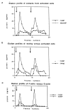
Fig. 1
Fig. 1
Elution profiles of F-actin, G-actin, and control (albumin) columns loaded with extracts from activated or resting platelets. In each case, the first arrow indicates the 0.5 mM Mg-ATP elution, and the second arrow indicates the subsequent 1.0 M KCl elution. A: Comparison of F-actin and control (BSA) columns loaded with extracts from ADP-activated platelets. The protein content of each fraction of 1.5 ml is plotted against the fraction number in each case. In this experiment, 30 ml of extract (1.2 mg/ml) was loaded onto two parallel columns, one a 3 ml F-actin (1 mg/ml actin) and the other a 3 ml control (albumin, 3 mg/ml) column. B: Comparison of resting versus activated platelet extracts chromatographed on F-actin columns. The profile for the ADP-extract eluting from F-actin column shown in A is compared to a profile from a parallel F-actin column loaded with extract from PGE-1 inhibited cells. The amount of protein in the extract from the PGE-1-inhibited cells was adjusted so that both columns were loaded with same amount of protein. C: Comparison of F-actin versus G-actin columns loaded with ADP-activated cell extracts. Twenty ml of soluble platelet protein at a concentration of 1 mg/ml from three units of platelet-rich plasma was loaded in parallel onto two columns: one, a 3 ml column of F-actin (2.5 mg/ml actin) and the other, a 3 ml column of G-actin (3.3 mg/ml actin). The protein concentration in each 1.5 ml fraction is plotted against the fraction number. Similar low levels of binding to the G-actin column were obtained for columns loaded with resting cell extracts.
Fig. 2
Fig. 2
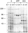
F-actin columns consistently enrich for particular proteins. Silver-stained 10% polyacrylamide gel of extracts and eluates from four different ADP-activated platelet extracts (X, lanes 1–4) run on four different 3 ml F-actin columns eluted with 0.5 mM Mg-ATP in column buffer (ATP, lanes 1–4) followed by 1.0 M KCl in the same buffer (KCl, lanes 1–4). Protein peaks were determined by Biorad Bradford protein assay, the peak fractions pooled and precipitated in 10% TCA and the pellet solubilized in SDS-containing gel sample buffer. One-tenth of the total elution peak from a 3 ml F-actin column was loaded onto each lane. S, molecular weight standards.
Fig. 3
Fig. 3
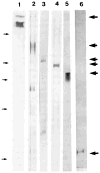
Elution of previously identified actin-binding proteins from F-actin columns. Western blots probed with (1) anti-macrophage actin-binding protein (filamin); (2) anti-glycoprotein IB; (3) anti-alpha-actinin; (4) anti-gelsolin; (5) anti-caldesmon; (6) anti-tropomyosin. Large arrows to the right indicate the migration of the antigens. Molecular weight standards indicated by small arrows on the left: myosin (200 kDa), phosphorylase a (98 kDa), albumin (68 kDa), ovalbumin (45 kDa), and carbonic anhydrase (29 kDa).
Fig. 4
Fig. 4
Western blots showing antigens recognized by antisera raised against gel-purified bands from F-actin column eluates. Antisera number is indicated at the top of each lane; C indicates a control blot that was incubated in parallel but without primary antibody. Antisera in A (C, 7, 16, 28, and 30) are blotted against whole platelet homogenates at 40 μg per lane, while antisera in B (6, 19, 32, and 33) are blotted against F-actin column eluates containing 8 μg per lane. Molecular weight standards are indicated by arrows to the left: myosin (200 kDa), phosphorylase a (98 kDa), albumin (68 kDa), and ovalbumin (45 kDa).
Fig. 5
Fig. 5
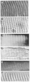
Platelet actin-binding proteins are found in the Z-band, and the A band in striated muscle. Frozen sections of human skeletal muscle stained with: (A) immunofluorescence of antiserum 7 and (B) same field, phase contrast; (C) antiserum 32; and (D) corresponding phase of the same field; (E) antiserum #16, and (F) corresponding phase image. Magnification: 2,400 X.
Fig. 6
Fig. 6
Staining patterns of platelet antigens in cultured fibroblasts compared to filamentous actin distribution by double label immunofluorescence. Double images of individual cells double-labeled with one of the antisera (left panels) and rhodamine phalloidin (right panels). Antisera are as indicated at the top of each set. Bar = 10 μm.
Fig. 6
Fig. 6
Staining patterns of platelet antigens in cultured fibroblasts compared to filamentous actin distribution by double label immunofluorescence. Double images of individual cells double-labeled with one of the antisera (left panels) and rhodamine phalloidin (right panels). Antisera are as indicated at the top of each set. Bar = 10 μm.
Fig. 7
Fig. 7
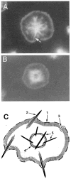
Four distinct actin domains in spread platelets. A and B: Two examples of platelets fixed and stained with rhodamine phalloidin 15 minutes after exposure to a glass coverslip. Arrow in A indicates a residual filopodial extension that has been engulfed by the advancing lamellipodium which surrounds the cell. In A the focal plane is on the glass surface; in B the focal plane is slightly above the glass revealing the contractile ring more clearly. C: An idealized schematic of the actin filament structures displayed in spread platelets: (1) leading edge; (2) filopodium; (3) lamellipodium; (4) contractile ring; (5) stress-like fibers. (In previous studies using electron microscopy, the cytoplasmic terminal of the filopodial actin bundles has been seen to connect with filaments that run the circumference of the cell [6, 21]. By immunofluorescence as described in this paper, that connection was not detected, and thus has been omitted from this diagram.)
Fig. 8
Fig. 8
Distribution of actin-binding proteins in discoid platelets and after activation. Discoid platelets (left column) stained with five different antisera as indicated to the left. Spread platelets fixed 15 min after exposure to glass coverslip and double-labeled by immunofluorescence with different antisera (middle column, antisera indicated to the far left) and with phalloidin (right column, same cell as in middle column). Arrows indicate filopodia (antiserum 21). Bar = 5 μm.

References

    1. Fox JEB, Philips DR. Polymerization and organization of actin filaments within platelets. Sem Hematol. 1983;20:243–260. - PubMed
    1. Fox JE. Regulation of platelet function by the cytoskeleton. Adv Exp Med Biol. 1993;344:175–185. - PubMed
    1. O’Halloran T, Beckerle MC, Burridge K. Identification of talin as a major cytoplasmic protein implicated in platelet activation. Nature (Lond) 1985;317:449–451. - PubMed
    1. Beckerle MC, Miller DE, Bertagnoli ME, Sarah Locke. Activation redistribution of the adhesion plaque protein, talin, in intact human platelets. J Cell Biol. 1989;109:3333–3346. - PMC - PubMed
    1. Nachmias VT, Golla R. Vinculin in relation to stress fibers in spread platelets. Cell Motil Cytoskel. 1991;20:190–202. - PubMed

Publication types

MeSH terms