. 1995;4(3):143-61.

Synthesis and maturation of viral transcripts in herpes simplex virus type 1 infected HeLa cells: the role of interchromatin granules

Affiliations

Synthesis and maturation of viral transcripts in herpes simplex virus type 1 infected HeLa cells: the role of interchromatin granules

S Besse et al. Gene Expr. 1995.

Abstract

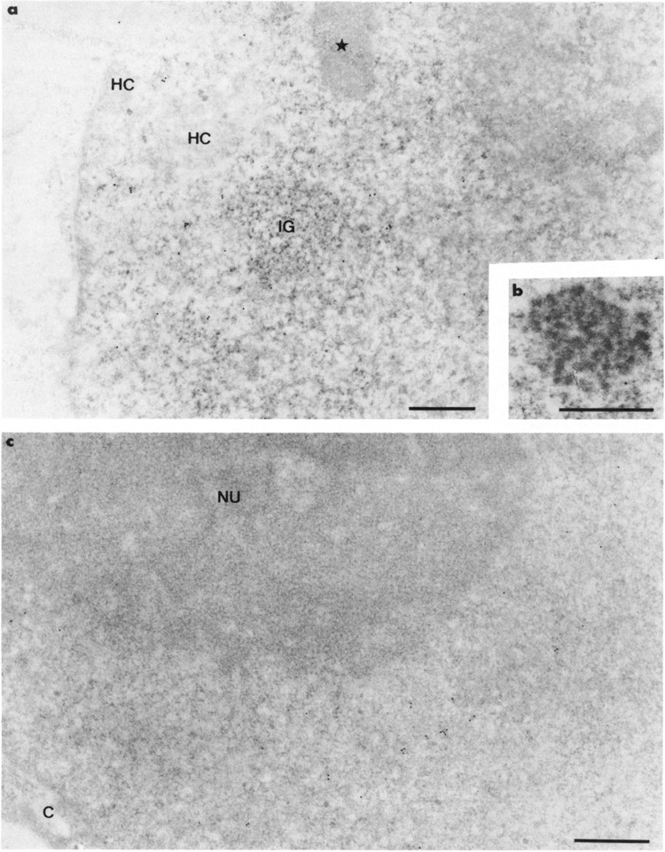
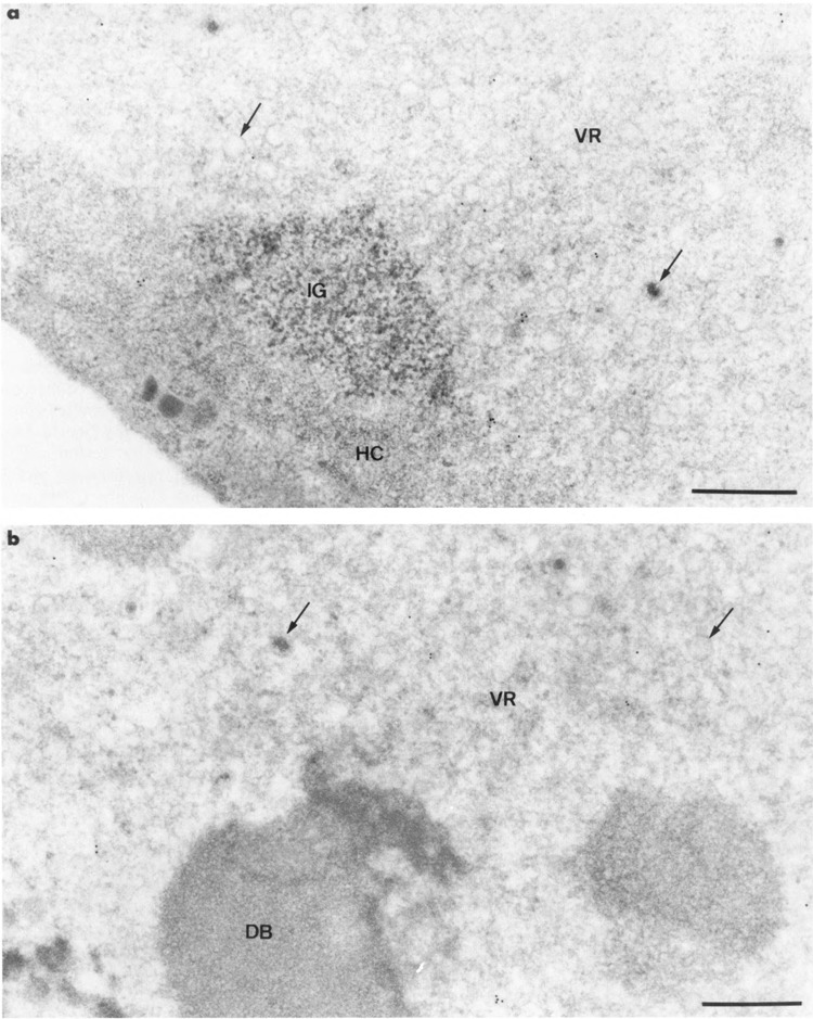
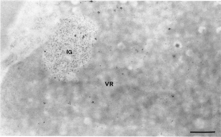
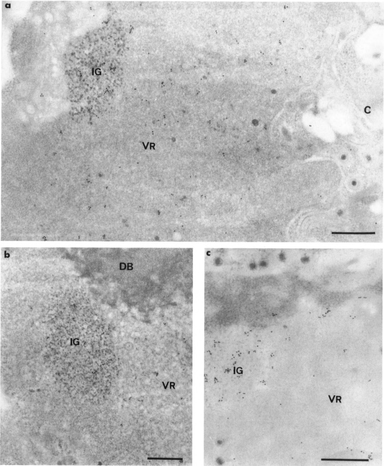
The response of the cellular RNA processing machinery to herpes simplex virus type 1 (HSV-1) infection was studied at the ultrastructural level in HeLa cells and compared to the distribution of RNA polymerase II molecules and viral RNA. Immunogold labeling of RNA polymerase II molecules revealed that viral genome transcription was restricted to filaments in an intranuclear, virus-induced region. This region also contained viral RNAs as revealed by in situ hybridization of two biotinylated viral DNA probes: a probe encompassing a limited portion of the viral genome (the F fragment) and a probe for the total genome. In addition, the latter probe revealed large amounts of viral RNA within the clusters of interchromatin granules, intranuclear structures of normal cells that became enlarged during HSV-1 infection. Components of spliceosomes were localized by in situ hybridization with biotinylated U1 and U2 DNA probes. The large viral region contained only traces of U1 and U2 RNAs, probably because of the low frequency of splices of viral transcripts. The clusters of interchromatin granules, however, accumulated U1 and U2 RNAs with the same frequency as in noninfected cells. Poly(A) RNA was detected by in situ hybridization of a biotinylated poly(dT) probe. Some was present over the filaments of the virus-induced region but most was accumulated in the clusters of interchromatin granules. Our data suggest, therefore, that the clusters of interchromatin granules, in addition to their involvement in spliceosome component assembly, might also be a transient storage site for some families of viral mRNA, possibly a sorting site that regulates their migration.


Figures

FIG. 1
FIG. 1
Intranuclear distribution of RNA polymerase II molecules in noninfected HeLa cells using 7C2 monoclonal antibody. Glutaraldehyde fixation and Lowicryl K4M embedding. Uranyl acetate staining. In (a), gold particles are present in the interchroma-tin space excluding the cluster of interchromatin granules (IG) and its associated zone (star). The condensed chromatin masses (HC) also are devoid of labeling. The coiled body in (b) and the nucleolus (NU) in (c) are entirely devoid of labeling. C: cytoplasm. Bars represent 0.5 μm.
FIG. 2
FIG. 2
Intranuclear distribution of RNA polymerase II molecules in HSV-1-infected HeLa cells using 7C2 monoclonal antibody. Formaldehyde fixation and Lowicryl K4M embedding. Uranyl acetate staining. Gold particles are present over the filaments of the large virus-induced region (VR) in (a) and (b) and absent over the viral capsids (arrows). The cluster of interchromatin granules (IG) and the marginated host chromatin (HC) in (a) and the intranuclear dense body (DB) in (b) are not labeled. Bars represent 0.5 μm.
FIG. 3
FIG. 3
Specificity of the 7C2 antibody. Increasing amounts of whole-cell extract (Manley et al., 1983) prepared from non-infected HeLa cells (10, 20, 40, and 80 μg in lanes 1–4, respectively) were loaded on a 10% polyacrylamide SDS gel (Laemmli, 1970). After electrophoresis, the proteins were transferred to a nitrocellulose filter, incubated with the 7C2 antibody, and protein/antibody complexes were revealed by the ECL Western blotting detection system (Amersham), as previously described (Chatton et al., 1994).
FIG. 4
FIG. 4
Intranuclear distribution of HSV-1 RNA in a HSV-1-infected HeLa cell by in situ hybridization of probe 2, a genomic probe. Formaldehyde fixation and Lowicryl K4M embedding. Protease-DNase pretreatment of sections prior to hybridization. Uranyl acetate staining. Gold particles are present over both the filaments of the virus-induced region (VR) and the cluster of interchromatin granules (IG). Bar represents 0.5 μm.
FIG. 5
FIG. 5
Intranuclear distribution of poly(A) RNA in HSV-1-infected cells by in situ hybridization of poly(dT) probe. Formaldehyde fixation and Lowicryl K4M embedding. Uranyl acetate staining. Comparison of the intensity of the labeling without (a) and with (b) protease digestion of section prior to hybridization. In the presence of the proteins of the section in (a), only a few gold particles are detectable over the viral region (VR) and the cluster of interchromatin granules (IG). Following the removal of the proteins of the section in (b), labeling is more intense, especially over the cluster of interchromatin granules (IG). The RNA-containing areas of the nucleolus, both fibrillar threads (F) and large, round masses of granules (G), are not labeled. Bars represent 0.5 μm.
FIG. 6
FIG. 6
Control: hybridization with poly(dT) probe following protease-RNase pretreatment of section. HSV-1-infected HeLa cell. Formaldehyde fixation and Lowicryl K4M embedding. Uranyl acetate staining. The viral region (VR) and the cluster of interchroma-tin granules (IG) are entirely devoid of labeling. HC: host chromatin; C: cytoplasm. Bar represents 0.5 μm.
FIG. 7
FIG. 7
Localization of U1 RNA in HSV-1-infected HeLa cells by in situ hybridization of an U1 DNA probe. Formaldehyde fixation and Lowicryl K4M embedding. Uranyl acetate staining. (a,b) Nondigested sections. Gold particles are numerous in the nucleus over the clusters of interchromatin granules (IG) and the virus-induced regions (VR). In (a), a few gold particles also are present in the cytoplasm (C) over its ribosome-rich areas whereas the viruses are devoid of labeling. In (b), the virus-induced intranuclear dense body (DB) is not labeled, (c) Protease pretreatment of section prior to hybridization. Elimination of the proteins of the section does not modify the intensity and the distribution of labeling. Gold particles are still associated with the intranuclear virus-induced region (VR) and the cluster of interchromatin granules (IG). Bars represent 0.5 μm.
FIG. 8
FIG. 8
Localization of U2 RNA by in situ hybridization of an U2 DNA probe. Formaldehyde fixation and Lowicryl K4M embedding. Uranyl acetate staining, (a) Noninfected HeLa cell. Gold particles are scattered over the interchromatin space and the clusters of interchromatin granules (IG). They are more numerous over the coiled body (CB) and totally absent over the interchromatin granule-associated zone (star), (b) HSV-1-infected HeLa cell. Gold particles are present over the cluster of interchromatin granules (IG) and, although at a lower extent, over the filaments of the viral region (VR). Arrow: viral capsid. Bars represent 0.5 μm.

References

    1. Bachellerie J. P., Puvion E., and Zalta J. P. (1975), Eur J Biochem 58, 327–337. - PubMed
    1. Bachenheimer S. L. and Roizman B. (1972), J Virol 10, 875–879. - PMC - PubMed
    1. Bachmann M., Falke D., Schröder H. C., and Müller W. E. (1989), J Gen Virol 70, 881–891. - PubMed
    1. Bartkoski M. J. Jr. and Roizman B. (1976), J Virol 20, 583–588. - PMC - PubMed
    1. Bartkoski M. J. Jr. and Roizman B. (1978), Virology 85, 146–156. - PubMed

Publication types

MeSH terms

LinkOut - more resources