Comparative Study
. 1995 Jan 15;3(1):63-78.
doi: 10.1016/s0969-2126(01)00135-6.

Structures of the native and swollen forms of cowpea chlorotic mottle virus determined by X-ray crystallography and cryo-electron microscopy

Affiliations
Comparative Study

Structures of the native and swollen forms of cowpea chlorotic mottle virus determined by X-ray crystallography and cryo-electron microscopy

J A Speir et al. Structure. .

Abstract

Background: RNA-protein interactions stabilize many viruses and also the nucleoprotein cores of enveloped animal viruses (e.g. retroviruses). The nucleoprotein particles are frequently pleomorphic and generally unstable due to the lack of strong protein-protein interactions in their capsids. Principles governing their structures are unknown because crystals of such nucleoprotein particles that diffract to high resolution have not previously been produced. Cowpea chlorotic mottle virions (CCMV) are typical of particles stabilized by RNA-protein interactions and it has been found that crystals that diffract beyond 4.5 A resolution are difficult to grow. However, we report here the purification of CCMV with an exceptionally mild procedure and the growth of crystals that diffract X-rays to 3.2 A resolution.

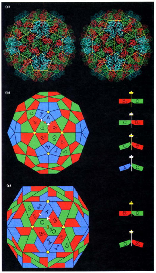
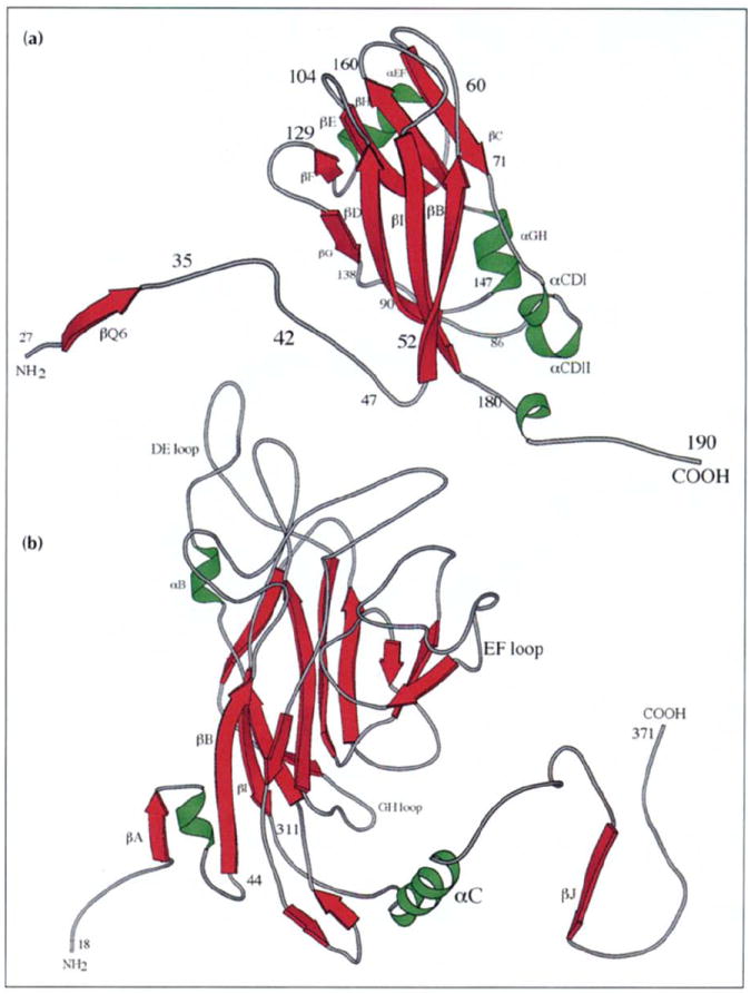
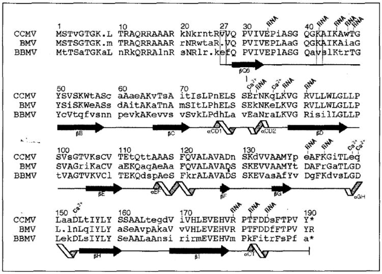
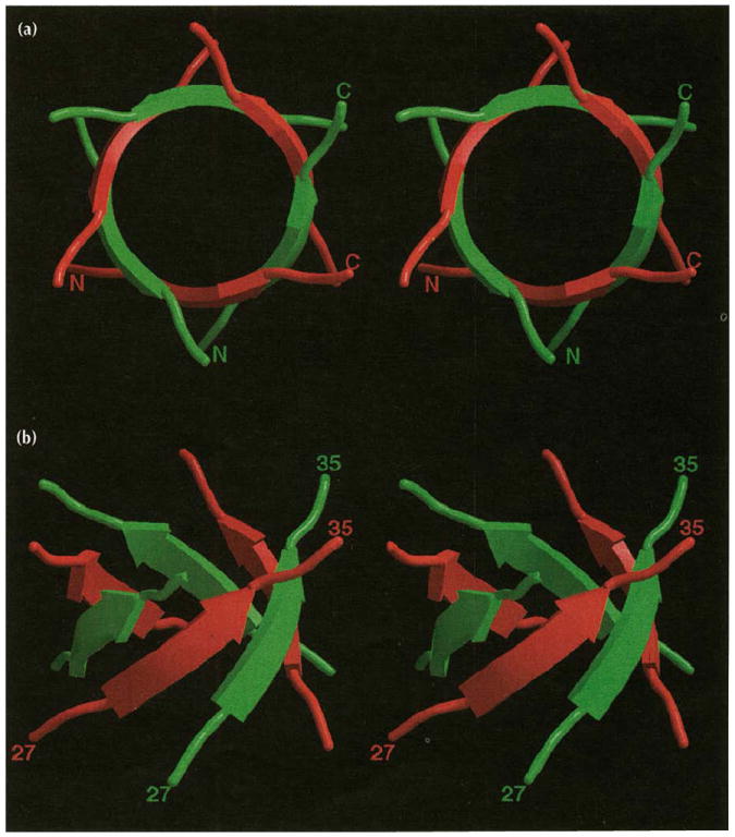
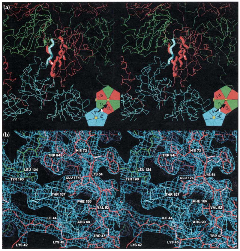
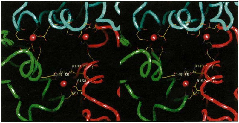
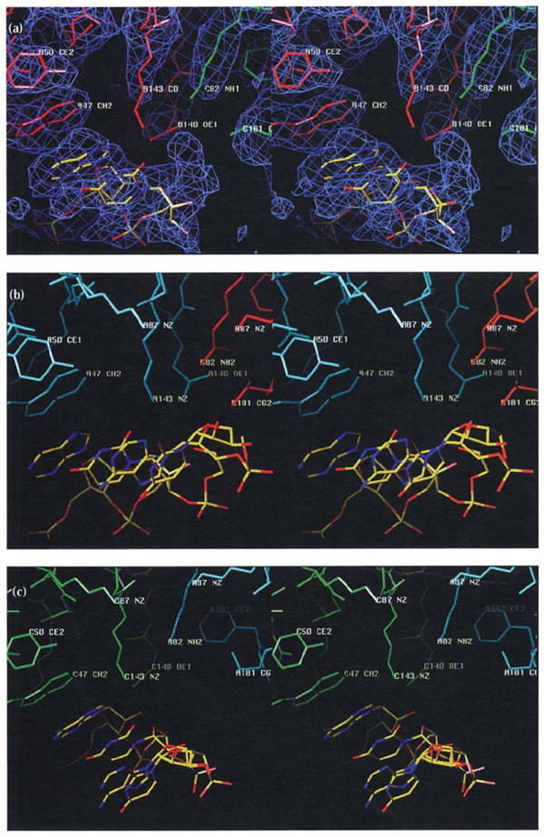
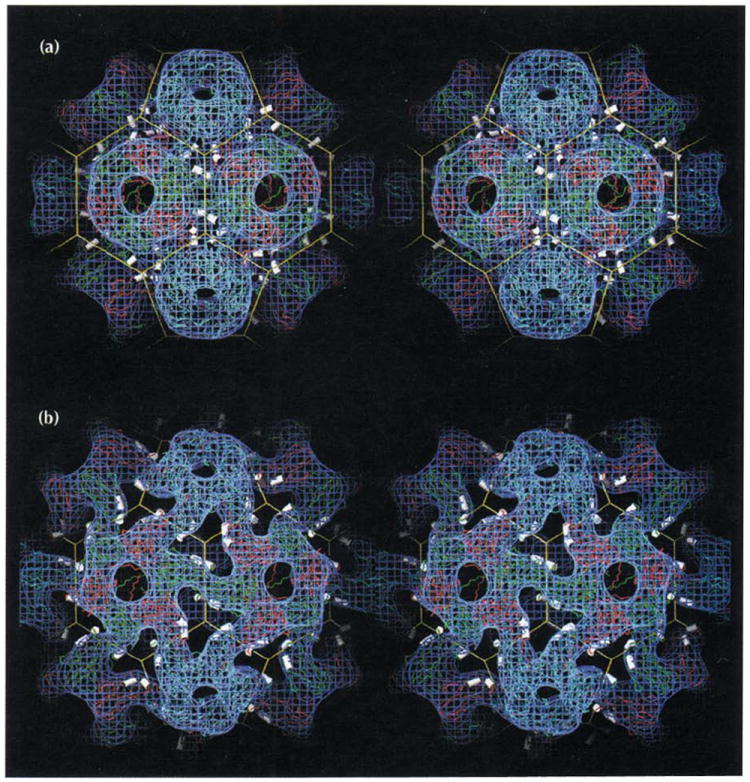
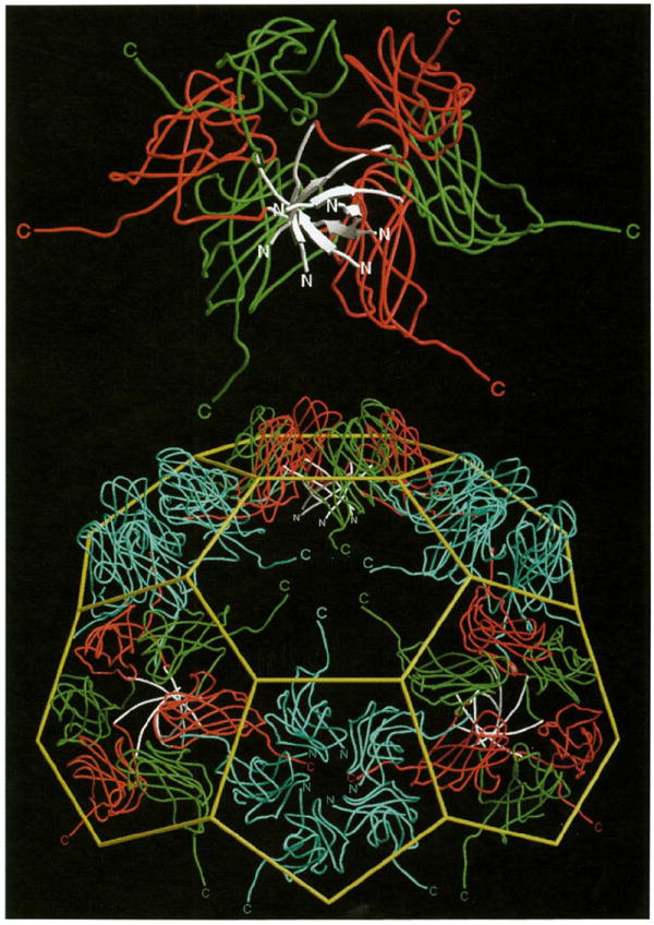
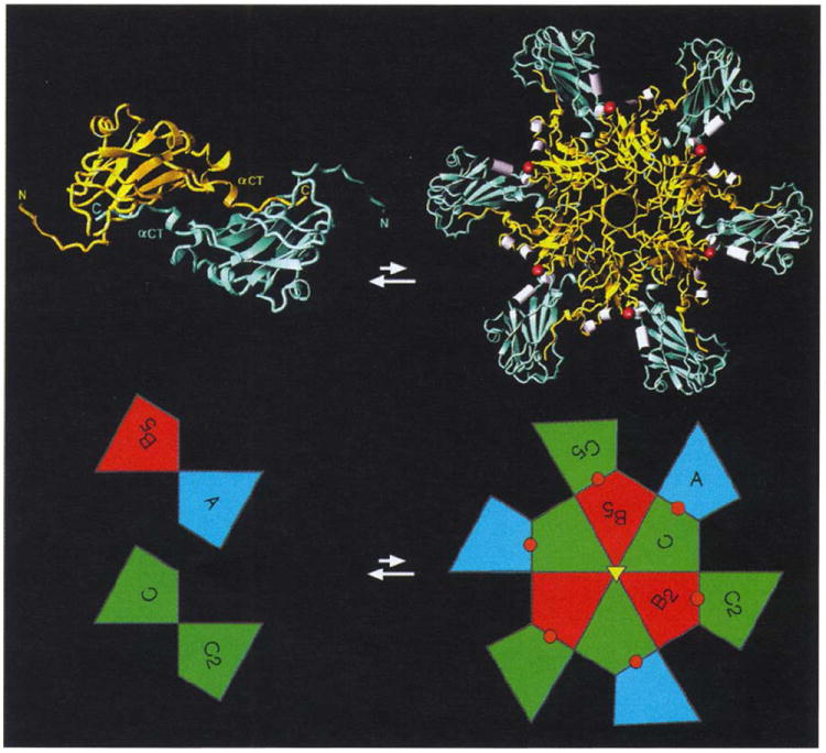
Results: The 3.2 A X-ray structure of native CCMV, an icosahedral (T = 3) RNA plant virus, shows novel quaternary structure interactions based on interwoven carboxyterminal polypeptides that extend from canonical capsid beta-barrel subunits. Additional particle stability is provided by intercapsomere contacts between metal ion mediated carboxyl cages and by protein interactions with regions of ordered RNA. The structure of a metal-free, swollen form of the virus was determined by cryo-electron microscopy and image reconstruction. Modeling of this structure with the X-ray coordinates of the native subunits shows that the 29 A radial expansion is due to electrostatic repulsion at the carboxyl cages and is stopped short of complete disassembly by preservation of interwoven carboxyl termini and protein-RNA contacts.

Conclusions: The CCMV capsid displays quaternary structural interactions that are unique compared with previously determined RNA virus structures. The loosely coupled hexamer and pentamer morphological units readily explain their versatile reassembly properties and the pH and metal ion dependent polymorphism observed in the virions. Association of capsomeres through inter-penetrating carboxy-terminal portions of the subunit polypeptides has been previously described only for the DNA tumor viruses, SV40 and polyoma.


Figures

Fig. 1
Fig. 1
Structure of the CCMV capsid and its novel geometrical features. (a) Stereoview of the protein shell as a Cα tracing. Color coding is defined in (b). The yellow cage represents the edges of a truncated icosahedron. (b) A truncated icosahedron model [38] shown in the same orientation as in (a). Positions of icosahedral rotation axes are marked by yellow symbols (pentagons: five-fold rotation axes; triangles: three-fold rotation axes; ovals: two-fold rotation axes). The central triangle with one five-fold (top) and two three-fold axes (lower left and right) at its vertices and containing polygons labeled A, B and C defines the icosahedral asymmetric unit. The polygons represent chemically identical protein subunits and within this area they occupy slightly different geometrical (chemical) environments and this is indicated by differences in their coloring. Polygons with subscripts are related to A, B and C by icosahedral symmetry (i.e. A to A5 by five-fold rotation). The apparent three-fold rotation axis at the center of the asymmetric unit Ivertices of cage in (a)] is not exact (quasi three-fold axis: white triangle) as it relates icosahedral threefold axes (quasi six-fold axes) to a five-fold axis outside of its local environment. Similarly, polygons labeled A and B5 form a quasi two-fold axis (white oval). Putative calcium-binding locations in one asymmetric unit of CCMV are marked by brown circles. Interactions between B2-C and between C-B5 polygons are defined by 180° dihedral angles (side view at top right) whereas bends similar in magnitude occur at the B-C and C-A polygon interfaces (138° and 142°, respectively) (side view at bottom right). (c) A rhombic triacontahedron model [38] shown in the same orientation and labeled identically to the truncated icosahedron model. The A, B and C polygons are co-planar within each asymmetric unit. Two such asymmetric units are co-planar by icosahedral two-fold symmetry giving the rhombic solid its prominent diamond shaped facets. The interaction between B2 and C remains planar (a 180° dihedral angle between two-fold related asymmetric units, top right) whereas the interaction of C and B5 adopts a 144° dihedral angle (bottom right). Hexamers are therefore best described as trimers of dimers.
Fig. 2
Fig. 2
Ribbon diagrams showing the tertiary structure of the (a) CCMV and (b) polyoma virus VP1 capsid subunits (VP1 drawn at smaller scale). Virus exteriors are at the top of each diagram. Selected residues and secondary structure elements are labeled. (Polyoma virus coordinates were kindly provided by Thilo Stehle and Steven Harrison, Harvard University.)
Fig. 3
Fig. 3
Sequence alignment of the bromovirus group capsid proteins and designation of the corresponding polypeptide secondary structure (CCMV, cowpea chlorotic mottle virus; BMV, bromegrass mosaic virus; BBMV, broad bean mosaic virus) [4,57,58]. Residue numbering at the top of each sequence block is for the CCMV protein with the left-most digit over the column to which the number corresponds. All other labels are based upon analysis of the high resolution CCMV capsid structure. Upper-case letters represent residues which are identical in at least two of the three sequences. The capsid proteins of CCMV and BMV share 70% identity whereas CCMV and BBMV are only 48% identical. Depending on the local capsid environment, ordered electron density for residues 27 (first box) to 190 (B and C subunits) or for residues 42 (second box) to 190 (A subunits) is observed. Residues which are involved in RNA binding and calcium ion coordination (Ca2+) are labeled. Secondary-structure elements determined by the program PROCHECK [59] are represented at the bottom of each sequence block. Extended black arrows and twisting ribbons represent β-sheet and α-helix secondary structure, respectively. These symbols are placed under the residues to which the represented structure has been assigned, and each labeled according to their location in the CCMV protein tertiary fold (Fig. 2a). Solid lines between β-sheets and α-helices have no regular secondary structure assigned. The dots indicate inserted gaps for purposes of alignment and the asterisks indicate that the equivalent terminal residues are not present in CCMV and BBMV.
Fig. 4
Fig. 4
Stereoviews of the β-hexamer shown as ribbon drawings that correspond to residues 27–35 of the B (red) and C (green) subunit amino termini. (a) The β-hexamer viewed directly down the quasi six-fold axis towards the particle interior. The amino and carboxyl termini are labeled for two of the six polypeptide renditions. Note that the strands labeled are not related by icosahedral symmetry. (b) The β-hexamer viewed approximately tangential to the virus surface. Residue numbers are given for the same polypeptides labeled above.
Fig. 5
Fig. 5
Stereoviews of the A-B5 quasi two-fold dimer contact responsible for binding pentamers to hexamers (see Fig. 1 for definitions and color coding). (a) An overview of the interaction showing the A subunits (blue) clustered around a five-fold axis and the B (red) and C (green) subunits clustered around a quasi six-fold axis. The bold tubes correspond to the regions of modeled electron density detailed in part (b). The inset is taken directly from Fig. 1b and displays the figure orientation relative to the particle symmetry axes. The black arrow represents the A subunit carboxyl terminus shown invading the B5 subunit here, and the visual orientation for (b). (b) A detailed view of the 3.2 Å electron density (light blue cage) for the interaction of the carboxy-terminal portion of the A subunit (blue wire model, residues 184–190) with the ‘clamp’ region of the B5 subunit (red wire model, residues 41–56, 91–95, 133–136, 171–176, the orientation is similar to that of the CCMV ribbon model in Fig. 2a). A small portion of the C subunit (green wire model, residues 123–125) is also visible. Hydrogen bonding of Thr187 by Glu174, the ‘fist in hand’ interactions of Phe186 (see text; Table 1), and the interaction of Leu124 of the quasi six-fold or five-fold related subunits with the Thr187 methyl group occur at each of the 180 clamp sites. This view is rotated 30° about the horizontal axis relative to (a). Views of the C-C2 icosahedral dimer contacts would be indistinguishable from those shown here except for the replacement of the pentamer with a second hexamer.
Fig. 6
Fig. 6
The putative quasi three-fold related calcium-binding sites in the CCMV capsid. The view is looking from the exterior to the interior of the virus directly down a quasi three-fold axis. Continuous tubes, color coded as in Fig. 1b, trace the polypeptide Cα atoms. Modeled calcium ions are displayed as brown spheres at each subunit interface (refer to Fig. 1b for their relative locations in the overall capsid) where only the potential calcium coordinating residues are shown, color coded by atom type (nitrogen, blue; oxygen, red; and carbon, yellow). Residues at the B-C subunit interface are labeled (as in Fig. 3 which shows the locations of the residues that interact with metal ions) in white. Positions of the αCDII and αGH helices are at the bottom edges of the quasi three-fold opening, and lining the sides of the opening more towards the exterior of the particle, respectively.
Fig. 7
Fig. 7
Similarly oriented stereoviews of the protein–RNA interactions at the A (blue), B (red), and C (green) subunit interfaces. The views are tangential to the protein shell looking away from the quasi three-fold axis along the subunit interfaces. The RNA model is colored according to atom type (nitrogen, blue; oxygen, red; and carbon, yellow). Selected residues are labeled as in Fig. 3, which shows the location of residues that interact with RNA. (a) The B-C subunit interface. Electron density (blue) at approximately 2σ contour level is shown for this interaction, but is left out of the following parts of the figure for clarity. (b) The A-B subunit interface. (c) The C-A subunit interface.
Fig. 8
Fig. 8
Stereoviews of two forms of the CCMV capsid showing that its polymorphism is controlled by changes in pH and metal ion concentrations. Atomic coordinates determined from X-ray crystallography and the truncated icosahedron cage are displayed as in Fig. 1a with electron density (blue) determined by cryo-electron microscopy. The helices which carry calcium-binding residues (αCDII and αGH in Figs 2a, 3, and 6) are displayed as white cylinders. (a) The CCMV capsid at pH 4.5. The atomic model is displayed as originally built in the X-ray electron density. The excellent fit between the 3.2 Å X-ray model and the 23 Å EM reconstruction shows the level of compatibility between the two independent structure determination techniques. Note the three pairs of CDII and GH helices positioned around each quasi three-fold axis (vertices of the truncated icosahedron). Calcium ions (180 per capsid) are bound between morphological units by residues associated with the helices (Table 2, Fig. 6). (b) The swollen CCMV capsid at pH 7.5 in the absence of metal ions determined at 28 Å resolution. Atomic coordinates from the high resolution native structure were modeled to fit the density from the cryoEM reconstruction.
Fig. 9
Fig. 9
An exterior view of a portion of the viral capsid close to a quasi six-fold axis. Continuous tubes represent subunit Cα tracings and are color coded as in Fig. 1. The yellow cage represents the idealized truncated icosahedron. Selected amino and carboxyl termini are labeled and color coded by subunit of origin. A hexameric morphological unit has been removed from the center of the contiguous shell and rotated about the horizontal axis to show on its underside the intracapsomere parallel β-structure (white) that stabilizes this morphological unit (the β-hexamer, Fig. 4). The hole left in the shell displays the disposition of residues 181–190 of the six carboxyl termini which invade the absent hexamer and interact with the clamp as shown in Fig. 5. Likewise, the removed hexamer displays the same carboxy-terminal residues that invade neighboring pentamers and hexamers pointing outward around its periphery. The orientation of the bottom pentamer provides a view down a five-fold axis. Note the lack of ordered interactions between the A subunit amino termini near the axis (i.e. disorder after residue Lys42), however, they have sufficient structure to clamp incoming B subunit carboxyl termini.
Fig. 10
Fig. 10
Proposed intermediates in CCMV assembly. A dimer (shown left and viewed from the exterior directly down a particle two-fold axis) and a 12mer (shown right and viewed from the particle exterior directly down a quasi six-fold axis) are displayed both as ribbon diagrams and as polygons from Fig. 1. Color coding of the ribbons is a mix of either B and C, or A and C colors (gold and light blue, respectively) and denotes those relationships, displayed by the polygon coloring, that cannot be distinguished until a greater portion of the capsid is formed. Amino termini and carboxyl termini are labeled for the dimer ribbon showing the exchange and clamping of carboxyl termini across both icosahedral and quasi two-fold axes (displayed in the lower part of the figure as C-C2 and A-B5 polygons). Six such dimers form a 12mer driven by the preferred formation of the β-hexamer. Helices that coordinate calcium (shown as red spheres in the ribbons and polygons) are displayed as white cylinders at subunit interfaces which are also capable of binding RNA.

References

    1. Speir A, Munshi S, Baker TS, Johnson JE. Preliminary X-ray data analysis of crystalline cowpea chlorotic mottle virus. Virology. 1993;193:234–241. - PubMed
    1. Francki RIB. The viruses and their taxonomy. In: Fraenkel-Conrat H, Wagner R, editors. The Plant Viruses. Plenum Press; New York: 1985. pp. 1–18.
    1. Lane LC. The bromoviruses. Adv Virus Res. 1974;19:151–220. - PubMed
    1. Dasgupta R, Kaesberg P. Complete nucleotide sequences of coat protein messenger RNAs of brome mosaic virus and cowpea chlorotic mottle virus. Nucleic Acids Res. 1982;10:703–713. - PMC - PubMed
    1. Baker TS. The packing of cowpea chlorotic mottle virus in crystalline monolayers. Proceedings of the Ninth International Congress on Electron Microscopy; 1978. pp. 24–25.

Publication types