

Peptide-induced infant status epilepticus causes neuronal death and synaptic reorganization
- PMID: 7756609
- PMCID: PMC3477862
- DOI: 10.1097/00001756-199501000-00013
Peptide-induced infant status epilepticus causes neuronal death and synaptic reorganization
Abstract
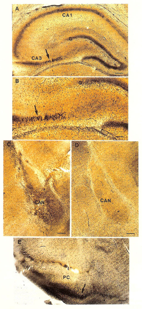
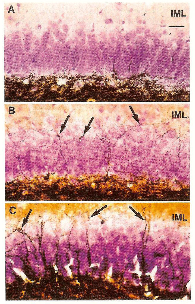
Status epilepticus (SE) produced by excitatory amino acids is a well established model in adult rodents. Limbic neuronal degeneration and synaptic reorganization observed after, for example, kainic acid-induced SE are considered relevant to human epilepsy. Kainic acid also produces severe seizures in infant rats, but neuronal injury and sprouting have not been demonstrated. The results of the present study show that corticotropin releasing hormone (CRH)-induced SE causes limbic neuronal death and reorganization in infant rats. In adults, CRH produced seizures at much higher doses, and no neuronal degeneration. As a modulator of the CNS stress response, CRH is activated in various 'stressful' circumstances. Its age-dependent ability to kill neurons represents a unique form of cell death potentially important in human medicine.
Figures
References
-
- Ben-Ari Y, Tremblay E, Riche D, et al. Neuroscience. 1981;6:1361–1391. - PubMed
-
- Lothman EW, Collins RC. Brain Res. 1981;218:299–318. - PubMed
-
- Cronin J, Dudek FE. Brain Res. 1988;474:181–184. - PubMed
-
- Nadler JW, Perry BW, Gentry C, et al. J Comp Neurol. 1981;196:549–569. - PubMed
-
- Cavazos JE, Sutula TP. Brain Res. 1990;527:1–6. - PubMed
Publication types
MeSH terms
Substances
Grants and funding
LinkOut - more resources
Full Text Sources
Research Materials