Increase of GPC levels in cultured mammalian cells during acidosis. A 31P MR spectroscopy study using a continuous bioreactor system
- PMID: 7760711
- PMCID: PMC10008650
- DOI: 10.1002/mrm.1910330317
Increase of GPC levels in cultured mammalian cells during acidosis. A 31P MR spectroscopy study using a continuous bioreactor system
Abstract
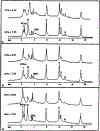
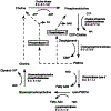
The purpose of this study was to study the metabolic events during a slow acidosis in three different cell lines by combining 31P magnetic resonance spectroscopy and hollow fiber bioreactor technology. The rate of change in intracellular pH, glycerophosphorylcholine (GPC), phophorylcholine (PCho), and nucleoside-triphosphate (NTP) levels were measured during 8 h of acidosis and 16 h of recovery in EPO, EAT, and RN1a cells, three cultured mammalians cell lines. Our results show a significant increase in GPC levels to 330 +/- 21.540 +/- 25, and 220 +/- 21% of their initial value correlated to a decrease of PCho levels to 57 +/- 14.58 +/- 17 and 45 +/- 15% of their initial value in EAT, RN1a, and EPO cells, respectively. These changes are discussed in terms of perturbation of energetic metabolism in cells undergoing a slow acidosis.
Figures


Similar articles
-
Phosphorous metabolites and steady-state energetics of transformed fibroblasts during three-dimensional growth.Am J Physiol Cell Physiol. 2002 Oct;283(4):C1287-97. doi: 10.1152/ajpcell.00097.2002. Am J Physiol Cell Physiol. 2002. PMID: 12225991
-
Nm23-transfected MDA-MB-435 human breast carcinoma cells form tumors with altered phospholipid metabolism and pH: a 31P nuclear magnetic resonance study in vivo and in vitro.Magn Reson Med. 1999 May;41(5):897-903. doi: 10.1002/(sici)1522-2594(199905)41:5<897::aid-mrm7>3.0.co;2-t. Magn Reson Med. 1999. PMID: 10332871
-
A comparison of in vivo and in vitro 31P NMR spectra from human breast tumours: variations in phospholipid metabolism.Br J Cancer. 1991 Apr;63(4):514-6. doi: 10.1038/bjc.1991.122. Br J Cancer. 1991. PMID: 2021535 Free PMC article.
-
Metabolic changes underlying 31P MR spectral alterations in human hepatic tumours.NMR Biomed. 1998 Nov;11(7):354-9. doi: 10.1002/(sici)1099-1492(1998110)11:7<354::aid-nbm515>3.0.co;2-n. NMR Biomed. 1998. PMID: 9859941 Review.
-
Glycerol phosphorylcholine (GPC) and serine ethanolamine phosphodiester (SEP): evolutionary mirrored metabolites and their potential metabolic roles.Comp Biochem Physiol Biochem Mol Biol. 1994 May;108(1):11-20. doi: 10.1016/0305-0491(94)90158-9. Comp Biochem Physiol Biochem Mol Biol. 1994. PMID: 8205386 Review.
Cited by
-
Phosphatidylcholine-Derived Lipid Mediators: The Crosstalk Between Cancer Cells and Immune Cells.Front Immunol. 2022 Feb 15;13:768606. doi: 10.3389/fimmu.2022.768606. eCollection 2022. Front Immunol. 2022. PMID: 35250970 Free PMC article. Review.
-
Microenvironmental Factors Modulating Tumor Lipid Metabolism: Paving the Way to Better Antitumoral Therapy.Front Oncol. 2021 Nov 23;11:777273. doi: 10.3389/fonc.2021.777273. eCollection 2021. Front Oncol. 2021. PMID: 34888248 Free PMC article. Review.
-
Human Melanoma-Cell Metabolic Profiling: Identification of Novel Biomarkers Indicating Metastasis.Int J Mol Sci. 2020 Mar 31;21(7):2436. doi: 10.3390/ijms21072436. Int J Mol Sci. 2020. PMID: 32244549 Free PMC article.
-
Serum Metabolomics Profiling Reveals Metabolic Alterations Prior to a Diagnosis with Non-Small Cell Lung Cancer among Chinese Community Residents: A Prospective Nested Case-Control Study.Metabolites. 2022 Sep 27;12(10):906. doi: 10.3390/metabo12100906. Metabolites. 2022. PMID: 36295809 Free PMC article.
-
Characterization of triple-negative breast cancer preclinical models provides functional evidence of metastatic progression.Int J Cancer. 2019 Oct 15;145(8):2267-2281. doi: 10.1002/ijc.32270. Epub 2019 Apr 2. Int J Cancer. 2019. PMID: 30860605 Free PMC article.
References
-
- Wike-Hooley JL, Haveman J, Reinhold HS, The relevance of tumor pH to the treatment of malignant disease. Radiother. Oncol 2, 343–366 (1984). - PubMed
-
- Gillies RJ, Liu Z, Bhujwalla Z, 31P-MRS measurements of extracellular pH of tumors using 3-aminopropylphosphonate. Am. J. Physiol 267, C195–C203 (1994). - PubMed
-
- Nunnally MH, Craig SW, Small changes in pH within the physiological range cause large changes in the consistency of actin-filamin mixtures. J. Cell. Biol 87, 218a (1980).
-
- Fitz JG, Trouillot TE, Scharschmidt BF, Effect of pH on membrane potential and K+ conductance in cultured rat hepatocytes. Am. J. Physiol 257, G961–G968 (1989). - PubMed
Publication types
MeSH terms
Substances
Grants and funding
LinkOut - more resources
Full Text Sources
Research Materials

