

Phosphorylation of CTP synthetase from Saccharomyces cerevisiae by protein kinase C
- PMID: 7797479
- PMCID: PMC1351267
- DOI: 10.1074/jbc.270.25.14983
Phosphorylation of CTP synthetase from Saccharomyces cerevisiae by protein kinase C
Abstract
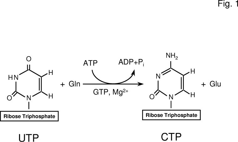
Phosphorylation of CTP synthetase (EC 6.3.4.2, UTP:ammonia ligase (ADP-forming)) from Saccharomyces cerevisiae protein kinase C was examined. Using pure CTP by synthetase as a substrate, protein kinase C activity was dose- and time-dependent and required calcium, diacylglycerol, and phosphatidylserine for full activation. Protein kinase C activity was also dependent on the concentration of CTP synthetase. Protein kinase C phosphorylated CTP synthetase on serine and threonine residues in vitro whereas the enzyme was primarily phosphorylated on serine residues in vivo. Phosphopeptide mapping analysis of CTP synthetase phosphorylated in vitro and in vivo indicated that the enzyme was phosphorylated on more than one site. Most of the phosphopeptides derived from CTP synthetase phosphorylated in vivo were the same as those derived from CTP synthetase phosphorylated by protein kinase C in vitro. The stoichiometry of the phosphorylation of native CTP synthetase was 0.4 mol of phosphate/mol of enzyme whereas the stoichiometry of the phosphorylation of alkaline phosphatase-treated CTP synthetase was 2.2 mol of phosphate/mol of enzyme. This indicated that CTP synthetase was purified in a phosphorylated state. Phosphorylation of CTP synthetase resulted in a 3-fold activation in enzyme activity whereas alkaline phosphatase treatment of CTP synthetase resulted in a 5-fold decrease in enzyme activity. Overall, the results reported here were consistent with the conclusion that CTP synthetase was regulated by protein kinase C phosphorylation.
Figures


Similar articles
-
Regulation of yeast CTP synthetase activity by protein kinase C.J Biol Chem. 1996 May 10;271(19):11113-9. doi: 10.1074/jbc.271.19.11113. J Biol Chem. 1996. PMID: 8626655
-
Phosphorylation and regulation of CTP synthetase from Saccharomyces cerevisiae by protein kinase A.J Biol Chem. 1996 Nov 15;271(46):28777-83. doi: 10.1074/jbc.271.46.28777. J Biol Chem. 1996. PMID: 8910520
-
Phosphorylation of human CTP synthetase 1 by protein kinase C: identification of Ser(462) and Thr(455) as major sites of phosphorylation.J Biol Chem. 2007 Jun 15;282(24):17613-22. doi: 10.1074/jbc.M702799200. Epub 2007 Apr 26. J Biol Chem. 2007. PMID: 17463002 Free PMC article.
-
CTP synthetase and its role in phospholipid synthesis in the yeast Saccharomyces cerevisiae.Prog Lipid Res. 2008 Sep;47(5):333-9. doi: 10.1016/j.plipres.2008.03.004. Epub 2008 Apr 7. Prog Lipid Res. 2008. PMID: 18439916 Free PMC article. Review.
-
Phospholipid synthesis in yeast: regulation by phosphorylation.Biochem Cell Biol. 2004 Feb;82(1):62-70. doi: 10.1139/o03-064. Biochem Cell Biol. 2004. PMID: 15052328 Review.
Cited by
-
Phosphorylation of phosphatidate phosphatase regulates its membrane association and physiological functions in Saccharomyces cerevisiae: identification of SER(602), THR(723), AND SER(744) as the sites phosphorylated by CDC28 (CDK1)-encoded cyclin-dependent kinase.J Biol Chem. 2011 Jan 14;286(2):1486-98. doi: 10.1074/jbc.M110.155598. Epub 2010 Nov 16. J Biol Chem. 2011. PMID: 21081492 Free PMC article.
-
Casein kinase II-mediated phosphorylation of lipin 1β phosphatidate phosphatase at Ser-285 and Ser-287 regulates its interaction with 14-3-3β protein.J Biol Chem. 2019 Feb 15;294(7):2365-2374. doi: 10.1074/jbc.RA118.007246. Epub 2019 Jan 7. J Biol Chem. 2019. PMID: 30617183 Free PMC article.
-
Phosphorylation of human CTP synthetase 1 by protein kinase A: identification of Thr455 as a major site of phosphorylation.J Biol Chem. 2007 Feb 23;282(8):5367-77. doi: 10.1074/jbc.M610993200. Epub 2006 Dec 22. J Biol Chem. 2007. PMID: 17189248 Free PMC article.
-
Phosphorylation of Yeast Pah1 Phosphatidate Phosphatase by Casein Kinase II Regulates Its Function in Lipid Metabolism.J Biol Chem. 2016 May 6;291(19):9974-90. doi: 10.1074/jbc.M116.726588. Epub 2016 Apr 4. J Biol Chem. 2016. PMID: 27044741 Free PMC article.
-
Pho85p-Pho80p phosphorylation of yeast Pah1p phosphatidate phosphatase regulates its activity, location, abundance, and function in lipid metabolism.J Biol Chem. 2012 Mar 30;287(14):11290-301. doi: 10.1074/jbc.M112.346023. Epub 2012 Feb 9. J Biol Chem. 2012. PMID: 22334681 Free PMC article.
References
Publication types
MeSH terms
Substances
Grants and funding
LinkOut - more resources
Full Text Sources
Molecular Biology Databases
