. 1994 Nov;130(1):95-105.
doi: 10.1006/exnr.1994.1188.

Morphine inhibits Purkinje cell survival and dendritic differentiation in organotypic cultures of the mouse cerebellum

Affiliations

Morphine inhibits Purkinje cell survival and dendritic differentiation in organotypic cultures of the mouse cerebellum

K F Hauser et al. Exp Neurol. 1994 Nov.

Abstract

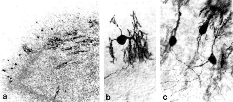
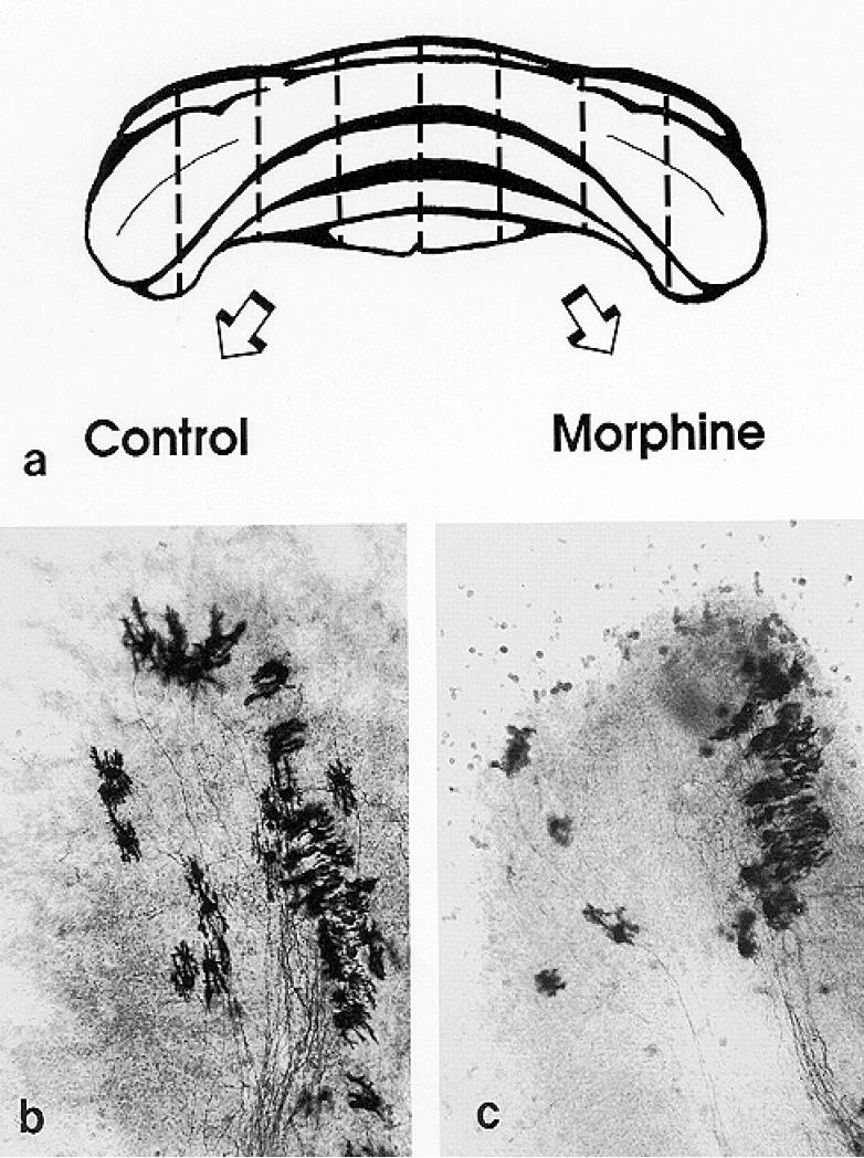
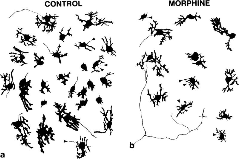
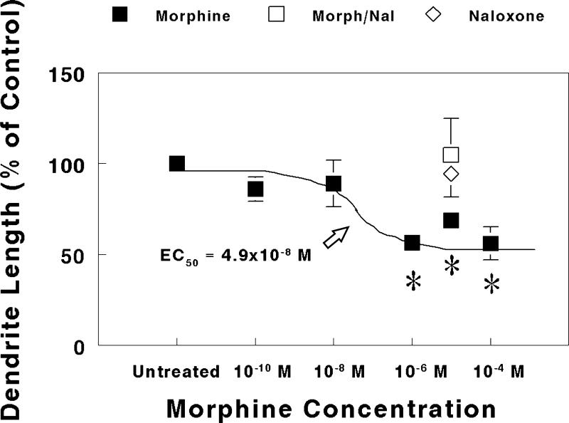
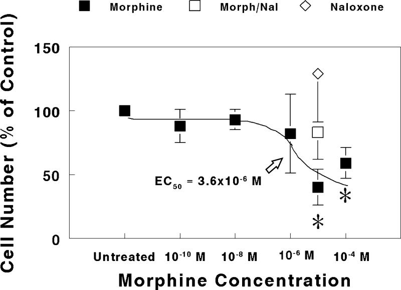
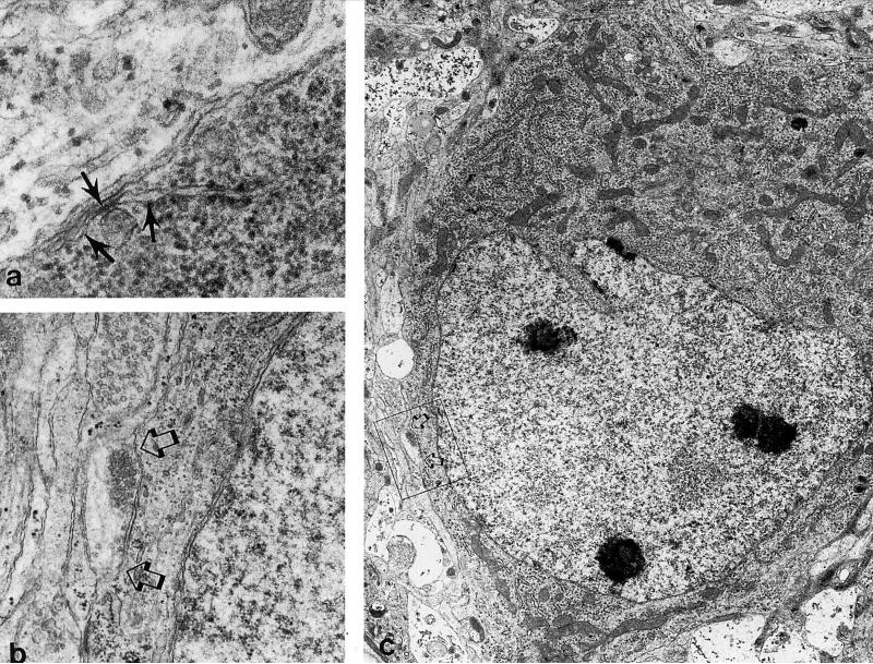
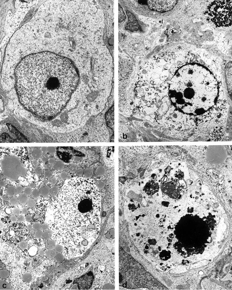
The effects of morphine on the morphogenesis and survival of calbindin-D28k-immunoreactive Purkinje cells were studied in organotypic explant cultures isolated from 1- or 7-day-old mouse cerebella. To reduce experimental variability, bilaterally matched pairs of organotypic cultures were used to compare the effects of opiate drug treatment. One explant within each pair was untreated, while the remaining explant was continuously treated for 7 to 10 days with morphine, morphine plus naloxone, or naloxone alone. In explants derived from 1-day-old mice, morphine treatment significantly reduced Purkinje cell dendritic length compared to symmetrically matched untreated control explants. The concentration of morphine estimated to cause a half-maximal reduction (EC50) in dendritic length was 4.9 x 10(-8) M. At higher concentrations (EC50 = 3.6 x 10(-6) M), morphine also significantly decreased the number of Purkinje cells in explants from 1-day-old mice compared to untreated explants. Electron microscopy identified increased numbers of degenerating Purkinje cells in explants derived from 1-day-old mice. This showed that high concentrations (10(-5) M) of morphine reduced Purkinje cell numbers by decreasing their rate of survival. In explants derived from 7-day-old mice, morphine (10(-5) M) neither affected Purkinje cell dendritic length nor cell numbers compared to symmetrically matched untreated (control) explants. Collectively, these findings suggest that morphine per se, through a direct action on the cerebellum, can affect Purkinje cell differentiation and survival. The results additionally suggest that there is a critical period during development when Purkinje cells are especially vulnerable to the effects of morphine.


Figures

FIG. 1
FIG. 1
Calbindin-D28k-immunoreactive Purkinje cells in cerebellar organotypic explant cultures. Bright-field photomicrographs of explants derived from 1 and 7-day-old mice at 7 to 10 days in vitro. (a). Dendritic size sometimes differed among Purkinje cells within the same explant (derived from a 1-day-old mouse). (b) Calbindin-D28k-immunoreactivity permitted visualization of Purkinje cell perikarya, dendrites and axons (explant derived from 1-day-old mouse). (c) Three relatively well differentiated Purkinje cells in an explant derived from a 7-day-old mouse. (a) 100 ×; (b & c) 480 ×.
FIG. 2
FIG. 2
Bilaterally-matched explant pairs were grown in the presence and absence of morphine. (a). Schematic drawing of the cerebellum from a 1-day-old mouse illustrating the “homologous- or mirror-pair” paradigm (63,64) used to assess the experimental effects of opiates. Tissue dissection was guided by existing anatomical divisions within the cerebellum. Cerebella from 1-day-old mice were divided into rostral and caudal portions at the primary fissure, and further divided into parasagittally-oriented explant cultures using additional cerebellar landmarks. Cerebella from 7-day-old mice are larger and are subdivided using different anatomical landmarks for dissection (see text). (b & c). Bilaterally-matched explant pairs have similar size, shape, and cytoarchitecture which is maintained in organotypic culture. For example, Purkinje cells in the untreated control explants (b) had larger dendrites compared to their matched-pair counterparts that were treated with 10-6 M morphine (c) at 7 to 10 days in vitro. (b & c) 220 ×.
FIG. 3
FIG. 3
Composite camera lucida drawings of calbindin-D28k-immunoreactive Purkinje cells illustrating the effects of high concentrations (10−5 M) of morphine on Purkinje cell numbers. Ccompared to untreated controls (a), continuous exposure to morphine (b) for 7 to 10 days in vitro caused concentration-dependent reductions in Purkinje cell numbers and dendritic length in cultures derived from 1-day-old mice (see Figs. 4 & 5). Numerous filopodial processes (arrowheads) were seen extending from the cell body or dendrites of calbindin-D28k-immunoreactive Purkinje cells. Filopodia are slender, thread-like processes of consistent diameter that transiently appear on growing neurons. Some filopodia terminate in a growth cones. (a & b) 320 ×.
FIG. 4
FIG. 4
Effect of increasing concentrations of morphine on Purkinje cell dendritic length in explants derived from 1-day-old mice. Purkinje cell dendritic length was measured and compared in bilaterally-matched paired-explant cultures. One explant within a matched-pair was given nutrient medium alone, while the other explant was continuously treated with morphine, morphine plus naloxone, or naloxone alone for 7 to 10 days in vitro. Note that concurrent 3 × 10⁻⁵ M naloxone plus 10−5 M morphine (Morph/Nal) treatment prevented morphine-induced deficits in dendritic length, while 3 × 10⁻⁵ M naloxone alone had no effect. Eight to 16 Purkinje cells were randomly sampled from each explant in a matched-pair. Determinations were made from explant pairs sampled from 6 mice for each drug concentration or treatment group. *P < 0.05 vs. untreated control.
FIG. 5
FIG. 5
Effect of increasing concentrations of morphine on Purkinje cell numbers in explants derived from 1-day-old mice. Purkinje cells were counted and their numbers compared in bilaterally-matched paired-explant cultures. One explant within a matched-pair was given nutrient medium alone, while the other explant was continuously treated with morphine, morphine plus naloxone, or naloxone alone for 7 to 10 days in vitro. Note that the reduction in cell numbers caused by 10-5 M morphine was prevented by concurrent treatment with 3 × 10⁻⁵ M naloxone (Morph/Nal), while naloxone (3 × 10⁻⁵ M) alone had no effect compared to untreated controls. Each determination was made from multiple matched-explant pairs sampled from 12 mice. *P < 0.05 vs. untreated control.
FIG. 6
FIG. 6
Electron micrographs of Purkinje cells in morphine-treated explants. Many Purkinje cells in morphine-treated explants show no morphologic signs of degeneration. (a) Subsurface or hypolemmal cisternae (arrows) are a characteristic cytoplasmic feature of Purkinje cells (47,55) (64,500×). (b) Detail of an axosomatic synapse (arrows in b & c) present on the Purkinje cell in (c) (19,100×). (c) Developing Purkinje cell (5,460×).
FIG. 7
FIG. 7
Degenerating cells in morphine-treated explants at 7 to 10 days in vitro. With progressive degeneration and loss of cellular morphology, it can be difficult to identify Purkinje cells with certainty. (a) A Purkinje cell with a deficit in cytoplasmic organelles and abnormally dense marginal heterochromatin (8,770×). (b) A dying cell surrounded by astrocytic processes containing numerous intermediate filaments (asterisks) (6,150×). (c) A degenerating cell with accumulated lipid and glycogen in the cytoplasm and partial destruction of the nuclear membrane. This cell had remnents of hypolemmal cisternae (not shown) (5,390×). (d) A large pyknotic cell positioned within the Purkinje cell layer that is difficult to identify with certainty (6,750×).

References

    1. ALTMAN J. Experimental reorganization of the cerebellar cortex. VII. Effects of late X-irradiation schedules that interfere with cell acquisition after stellate cells are formed. J.Comp.Neurol. 1976;165:65–76. - PubMed
    1. BAPTISTA CA, HATTEN ME, BLAZESKI R, MASON CA. Cell-cell interactions influence survival and differentiation of purified Purkinje cells in vitro. Neuron. 1994;12:243–260. - PubMed
    1. BAYER SA, ALTMAN J, RUSSO RJ, ZHANG X. Timetables of neurogenesis in the human brain based on experimentally determined patterns in the rat. Neurotoxicol. 1993;14:83–144. - PubMed
    1. BHARGAVA HN, VILLAR VM, RAHMANI NH, LARSEN AK. Time course of the distribution of morphine in brain regions, spinal cord and serum following intravenous injection to rats of differing ages. Pharmacology. 1993;47:13–23. - PubMed
    1. BRUNI JF, VANVUGT D, MARSHALL S, MEITES J. Effects of naloxone, morphine and methionine enkephalin on serum prolactin, luteinizing hormone, follicle stimulating hormone, thyroid stimulating hormone and growth hormone. Life Sci. 1977;21:461. - PubMed

Publication types

LinkOut - more resources