. 1994;4(1-2):19-28.

Structure and expression of a polyubiquitin gene from the crustacean Artemia

Affiliations

Structure and expression of a polyubiquitin gene from the crustacean Artemia

E Franco et al. Gene Expr. 1994.

Abstract

We have characterized two polyubiquitin genes from the crustacean Artemia franciscana. One of them, Ubi1, has nine ubiquitin units and an intron of a minimum size of 3.5 kb that ends 7 bp before the initiator ATG. The 5' end of the transcript from this gene has been identified by anchored PCR. The existence of the other gene (Ubi2) was inferred from several cDNA clones that differ from Ubi1 in the C-terminal extension and in the 3' untranslated region as well as in the nucleotide sequence of the coding region. We find two transcripts of ubiquitin genes, of 2.7 and 3.3 kb. Hybridization of RNA blots with an oligonucleotide specific for Ubi2 gene demonstrates that this gene codes for the 3.3 kb transcript. Ubiquitin messenger RNAs are present in the dormant embryos and their steady-state levels are maximum at 8 h after resumption of development, declining thereafter. The Ubi2 gene transcripts are less abundant but its proportion in relation to the other transcript does not vary with development.


Figures

FIG 1
FIG 1
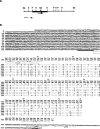
Sequence and structure of the plasmid pcUbq. The sequence is aligned to show the ubiquitin units. Similar bases are denoted by a dash. The stop codon is marked with three asterisks. The oligonucleotides derived from pcUbq described in the Materials and Methods section are indicated below the sequence. The ATTAAA polyadenylation signal is underlined.
FIG 2
FIG 2
Structure of the Ubil gene. (A) Restriction map of λgArtU5. The sequenced fragments are shown as a solid line. The arrow indicates the 5′ → 3′ direction of the gene. Restriction enzymes are: E, EcoRI; H, HindIII; P, Pst I; S, SalI. (B) Sequence and structure of the Ubil gene. Conventions are the same as in Fig. 1. The intron acceptor site is double underlined and the ATTAAA polyadenylation signal is underlined. Oligonucleotides described in the Materials and Methods section are also indicated.
FIG 3
FIG 3
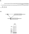
Anchored PCR. (A) Sequence of the fragment obtained as described in the Materials and Methods section and in the text. The oligonucleotide derived from this sequence is indicated. (B) Diagram showing the setup of the experiment to demonstrate that the intron is processed. (C) Results of the experiment. RNA (1, 3, and 5 μg) from 8 h of development was used to perform a reverse transcription-linked PCR, as described in the text. The products of the PCR were electrophoresed in 3% agarose gel and transferred to a nitrocellulose membrane. The membrane was hybridized with oligonucleotide C-l. Molecular weight markers (in kb) pUC19 plasmid digested with HpaII and TaqI.
FIG 4
FIG 4
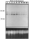
Hybridization of RNAs from different times of development with an ubiquitin-coding sequence probe; 15 μg of total RNA was used per lane. Markers are ribosomal RNAs from the yeast Saccharomyces cerevisiae. The lower panel shows the ethidium bromide-stained gel. In this experiment, hatching of the nauplii was at 18 h of development.
FIG 5
FIG 5
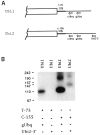
Expression of Ubi1 and Ubi2 genes. (A) Diagram of the 3′ regions of Ubi1 and Ubi2 genes, showing the locations of the sense and antisense primers used. (B) cDNA made from RNA from 12 h of development was amplified with the primers indicated under each lane, separated by gel electrophoresis in 3% agar-ose, blotted, and hybridized with cUbq oligonucleotide. Molecular weight markers (in bp) are derived from pBlueskript plasmid digested with HpaII.
FIG 6
FIG 6
Identification of Ubi2 mRNA as the 3.3 kb band. Poly (A) + RNA obtained from 25 h of development (15 μg) was electrophoresed and hybridized sequentially to (A), the 130 bp fragment of the 5′ end obtained by anchored PCR, (B) the 3′ end 137 bp fragment of pcUbq, and (C), the Ubi2-3′ oligonucleotide.

Similar articles

Cited by

References

    1. Baker R. T. and Bord P. G. (1987), Nucleic Acids Res 15, 443–463. - PMC - PubMed
    1. Bond U. and Schlesinger M. J. (1986), Mol Cell Biol 6, 4602–4610. - PMC - PubMed
    1. Browne R. A., Sorgeloos P., and Trotman C. N. A. (1991), Artemia Biology, CRC Press, Boca Raton, FL.
    1. Busch H. and Goldknopf L. L. (1981), Mol Cell Biochem 40, 173–187. - PubMed
    1. Cabrera y Poch H. L., Arribas C., and Izquierdo M. (1990), Nucleic Acids Res 18, 3994. - PMC - PubMed

Publication types

Associated data

LinkOut - more resources