Acute herpetic esophagitis--a case report
- PMID: 7865487
- PMCID: PMC4532066
- DOI: 10.3904/kjim.1994.9.2.120
Acute herpetic esophagitis--a case report
Abstract
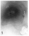
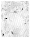
We report a case of acute herpetic esophagitis in a 33 year old man who was presumed to be immuno-compromised following prolonged steroid and cyclosporin treatment for acute rejection of a transplanted kidney. In Korea, all reported cases of herpetic esophagitis have been diagnosed in immuno-compromised and debilitated patients with a typical endoscopic appearance of ulcerating lesions. However, our patient showed multiple vesicular lesions without ulcer along the entire esophagus. The diagnosis was confirmed by colorimetric detection of herpes virus DNA using in situ hybridization. The endoscopic findings reported herein probably represent the typical early stage of acute herpetic esophagitis.
Figures


Similar articles
-
Herpes simplex virus esophagitis in the immunocompetent host: an overview.Am J Gastroenterol. 2000 Sep;95(9):2171-6. doi: 10.1111/j.1572-0241.2000.02299.x. Am J Gastroenterol. 2000. PMID: 11007213 Review.
-
[Diagnosis of herpetic esophagitis in the immunocompetent subject by PCR (Herpès Consensus Générique-Argène). Report of six cases].Pathol Biol (Paris). 2009 Feb;57(1):101-6. doi: 10.1016/j.patbio.2008.07.005. Epub 2008 Oct 7. Pathol Biol (Paris). 2009. PMID: 18842356 French.
-
[Herpetic esophagitis in 5 immunocompetent patients].Ned Tijdschr Geneeskd. 1996 Jun 29;140(26):1367-71. Ned Tijdschr Geneeskd. 1996. PMID: 8710027 Dutch.
-
Epigastric pain associated with herpes esophagitis: case report.BMC Infect Dis. 2020 Oct 14;20(1):754. doi: 10.1186/s12879-020-05487-5. BMC Infect Dis. 2020. PMID: 33054791 Free PMC article.
-
Herpes esophagitis in healthy adults and adolescents: report of 3 cases and review of the literature.Medicine (Baltimore). 2010 Jul;89(4):204-210. doi: 10.1097/MD.0b013e3181e949ed. Medicine (Baltimore). 2010. PMID: 20616659 Review.
Cited by
-
Seronegative Herpes simplex Associated Esophagogastric Ulcer after Liver Transplantation.Case Rep Gastroenterol. 2008 Mar 13;2(1):103-8. doi: 10.1159/000119113. Case Rep Gastroenterol. 2008. PMID: 21490847 Free PMC article.
References
-
- Berg JW. Esophageal herpes: A complication of cancer therapy. Cancer. 1955;8:731. - PubMed
-
- Agha FP, Lee HH, Nostant TT. Herpetic esophagitis: A diagnostic challenge in immunocompromised patients. Am J Gastroenterol. 1986;81:246. - PubMed
-
- Buss DH, Scharyi M. Herpes virus infection of the esophagus and other visceral organs in adults. Am J Med. 1979;66:457. - PubMed
-
- Depew WT, Prentice RSA, Beck IT, Blakeman JM, Dacosta LA. Herpes simplex ulcerative esophagitis in healthy patients. Am J Gastroenterol. 1977;68:381. - PubMed
-
- Deshmukh M, Shah R, McCallum RW. Experience of herpes esophagitis in otherwise healthy patients. Am J Gastroenterol. 1984;79:173. - PubMed
Publication types
MeSH terms
Substances
LinkOut - more resources
Full Text Sources
Medical
