. 1994 Nov;63(5):1872-9.
doi: 10.1046/j.1471-4159.1994.63051872.x.

Increased neuronal beta-amyloid precursor protein expression in human temporal lobe epilepsy: association with interleukin-1 alpha immunoreactivity

Affiliations

Increased neuronal beta-amyloid precursor protein expression in human temporal lobe epilepsy: association with interleukin-1 alpha immunoreactivity

J G Sheng et al. J Neurochem. 1994 Nov.

Abstract

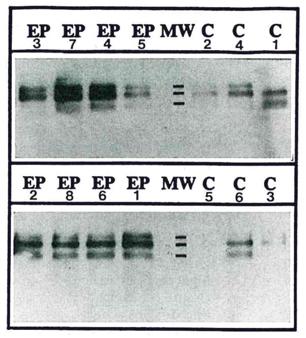
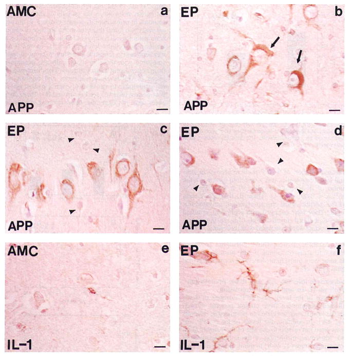
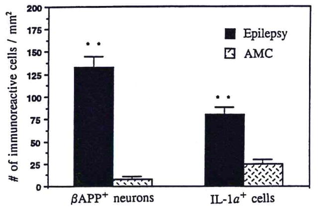
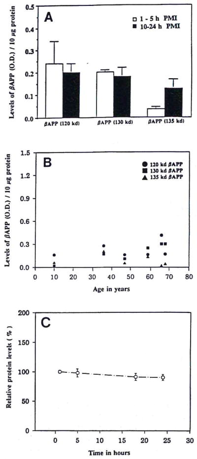
Levels of immunoreactive beta-amyloid precursor protein and interleukin-1 alpha were found to be elevated in surgically resected human temporal lobe tissue from patients with intractable epilepsy compared with postmortem tissue from neurologically unaffected patients (controls). In tissue from epileptics, the levels of the 135-kDa beta-amyloid precursor protein isoform were elevated to fourfold (p < 0.05) those of controls and those of the 130-kDa isoform to threefold (p < 0.05), whereas those of the 120-kDa isoform (p > 0.05) were not different from control values. beta-Amyloid precursor protein-immunoreactive neurons were 16 times more numerous, and their cytoplasm and proximal processes were more intensely immunoreactive in tissue sections from epileptics than controls (133 +/- 12 vs. 8 +/- 3/mm2; p < 0.001). However, neither beta-amyloid precursor protein-immunoreactive dystrophic neurites nor beta-amyloid deposits were found in this tissue. Interleukin-1 alpha-immunoreactive cells (microglia) were three times more numerous in epileptics than in controls (80 +/- 8 vs. 25 +/- 5/mm2; p < 0.001), and these cells were often found adjacent to beta-amyloid precursor protein-immunoreactive neuronal cell bodies. Our findings, together with functions established in vitro for interleukin-1, suggest that increased expression of this protein contributes to the increased levels of beta-amyloid precursor protein in epileptics, thus indicating a potential role for both of these proteins in the neuronal dysfunctions, e.g., hyperexcitability, characteristic of epilepsy.


Figures

FIG. 1
FIG. 1
Western immunoblots depict β-APP-immunoreactive products in samples of resected temporal lobes from eight epileptic patients (EP) and in samples from analogous regions of temporal lobe collected postmortem from six controls (C). Monoclonal anti-β-APP antibody (clone 22C11) sensitively labeled three bands on western immunoblots at molecular weight (MW) marks shown at 135K, 130K, and 120K.
FIG. 2
FIG. 2
Photomicrographs of β-APP (APP; a–d) and IL-1α (IL-1; e and f) immunoreaction product in cells in tissue sections of temporal lobe from epileptics (EP; b–d and f) and controls (AMC; a and e). Simultaneous immunohistochemical reaction with anti-β-APP or anti-IL-1α antibodies was performed on 10-μm-thick paraffin-embedded temporal lobe tissue sections from epileptics and controls, as described in Materials and Methods, a: β-APP-immunoreacted tissue sections of temporal lobe gray matter from control (AMC) illustrate the paucity of immunoreaction product in neurons (layer V). b: β-APP-positive neurons in analogous sections of temporal lobe (layer V) from an epileptic (EP 7) demonstrate the relatively high levels of immunoreaction product (brown); β-APP-positive intracellular tangle-like structures were observed in some neurons (arrow), c and d: β-APP-positive neurons in tissue sections from case EP 3 show large pyramidal neurons (c) in temporal lobe cortical layer V, containing high levels of β-APP immunoreaction product (brown) relative to the levels in small and medium neurons (c and d; arrowheads). Some small neurons in layer VI in case EP 3 (d) also contained high levels of β-APP immunoreaction product (brown), whereas others did not (arrowhead), e: Tissue section shows the very low IL-1α immunoreactivity characteristic of cells in gray matter of controls (AMC). f: Tissue section from an epileptic (EP 7) illustrates the increase in the number, size, and immunoreactive intensity of IL-1α-positive cells (brown) in epilepsy; many were found immediately adjacent to neurons in layer V of temporal lobe. Bar = 15 μm.
FIG. 3
FIG. 3
Bar graph compares the number of β-APP-positive neurons/mm2 and IL-1α-positive cells/mm2 in gray matter of temporal lobe from epileptic patients and controls (AMC). β-APP-positive neurons and IL-1α-positive cells were counted, as described in Materials and Methods, in tissue sections from each of eight epileptic and six control patients. The data are mean ± SEM (bars) values. **p < 0.001.
FIG. 4
FIG. 4
A: Bar graph correlates the levels of three β-APP isoforms in temporal lobe samples with postmortem intervals (PMIs) of control patients. Data are mean ± SEM (bars) values for three patients in each group. B: Dot graph correlates the levels of three β-APP isoforms in temporal lobe samples with ages of control patients. C: Loss of measurable protein in homogenates of surgical temporal lobe samples of epileptic patients incubated at room temperature between 1 and 24 h. Data are mean ± SEM (bars) values for six samples relative to the initial (1-h) value.

Similar articles

Cited by

References

    1. Arai H, Lee VM, Otvos L, Jr, Greenberg BD, Lowery DE, Sharma SK, Schmidt ML, Trojanowski JQ. Defined neurofilament, tau and β-amyloid precursor protein epitopes distinguish Alzheimer from non-Alzheimer senile plaque. Proc Natl Acad Sci USA. 1990;87:2249–2253. - PMC - PubMed
    1. Araki W, Kitaguchi N, Tokushima Y, Ishii K, Aratake H, Shimohama S, Nakamura S, Kimura J. Trophic effect of β-amyloid precursor protein on cerebral cortical neurons in culture. Biochem Biophys Res Commun. 1991;181:265–271. - PubMed
    1. Armstrong DD. The neuropathology of temporal lobe epilepsy. J Neuropathol Exp Neurol. 1993;52:433–443. - PubMed
    1. Ban EM, Sarlieve LL, Haour FG. Interleukin-1 binding sites on astrocytes. Neuroscience. 1993;52:725–733. - PubMed
    1. Bancher C, Brunner C, Lassmann H, Budka H, Jellinger K, Wiche G, Seitelberger F, Grundke-Iqbal I, Iqbal K, Wisniewski HM. Accumulation of abnormally phosphorylated τ precedes the formation of neurofibrillary tangles in Alzheimer’s disease. Brain Res. 1989;477:90–99. - PubMed

Publication types

MeSH terms

Substances

LinkOut - more resources