. 1994 Dec 2;79(5):779-90.
doi: 10.1016/0092-8674(94)90068-x.

Xenopus chordin: a novel dorsalizing factor activated by organizer-specific homeobox genes

Affiliations

Xenopus chordin: a novel dorsalizing factor activated by organizer-specific homeobox genes

Y Sasai et al. Cell. .

Abstract

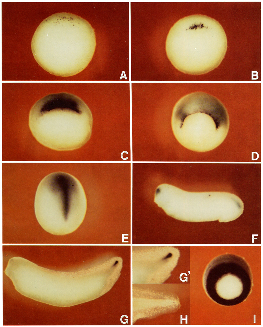
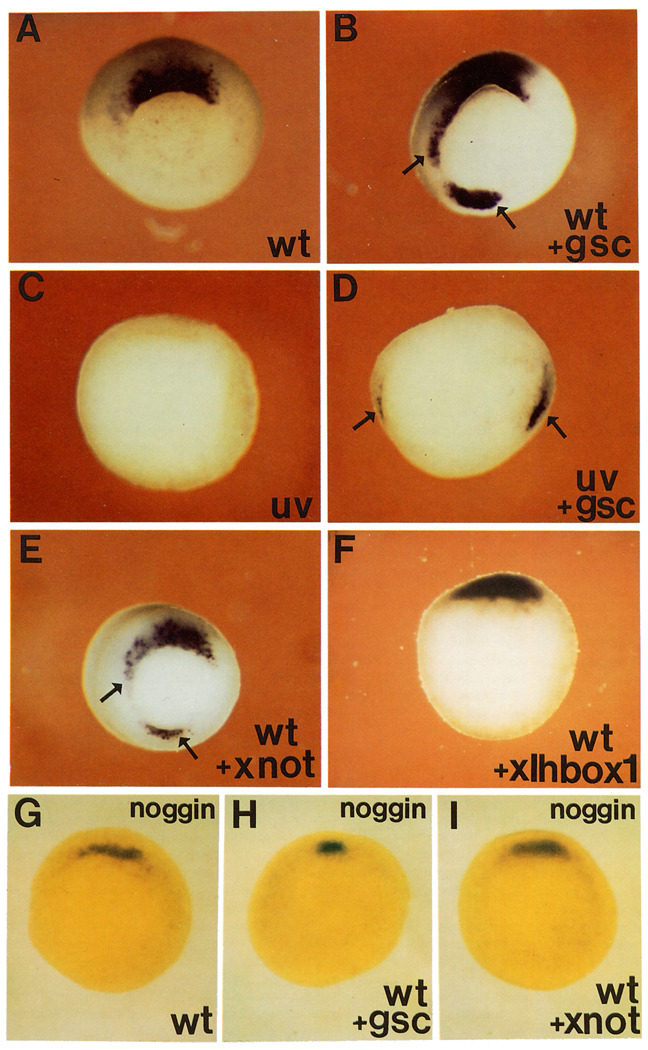
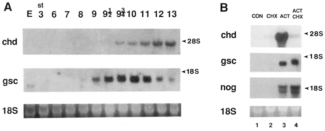
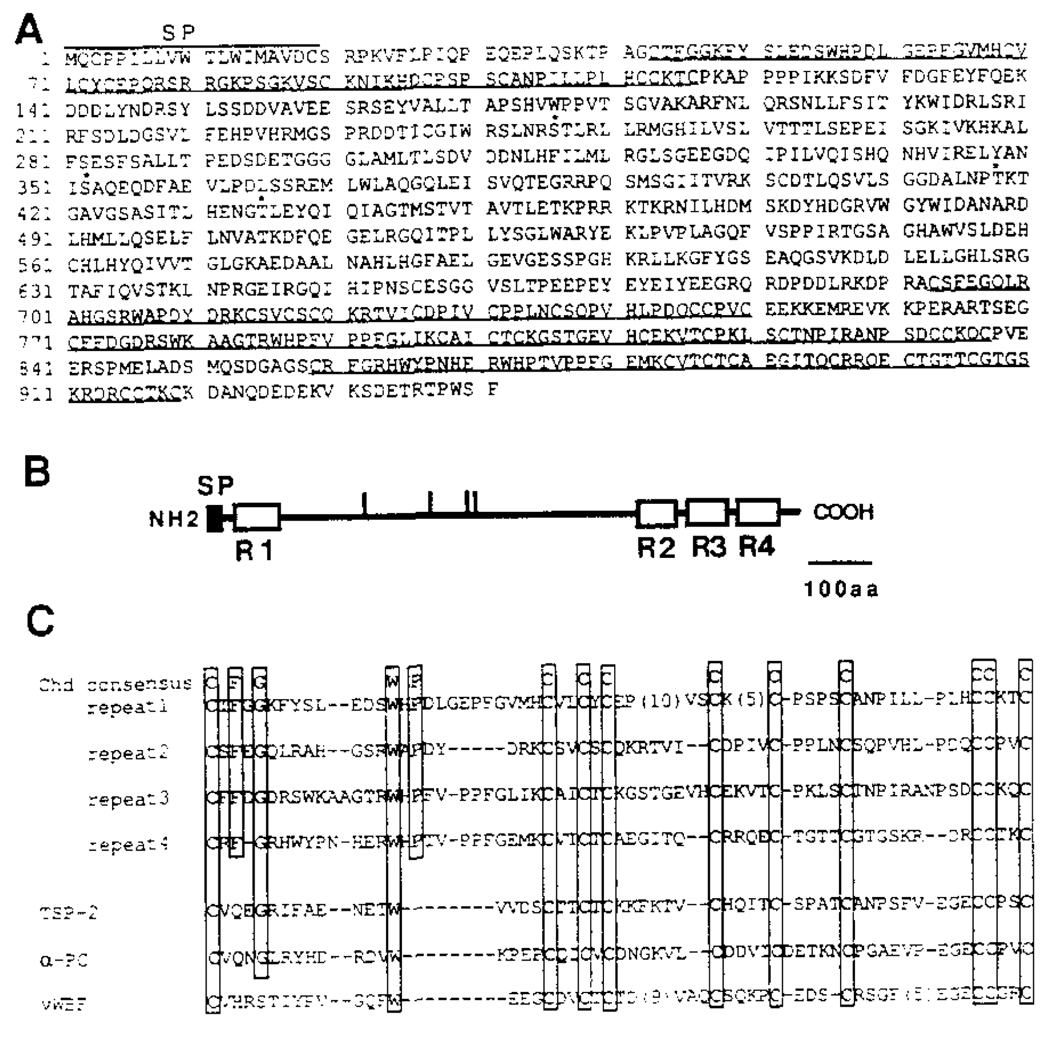
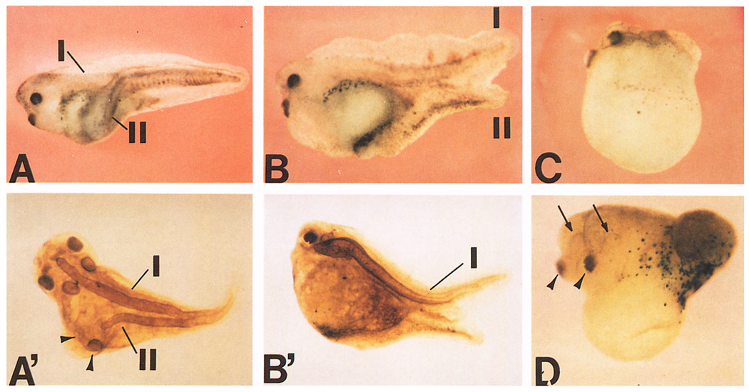
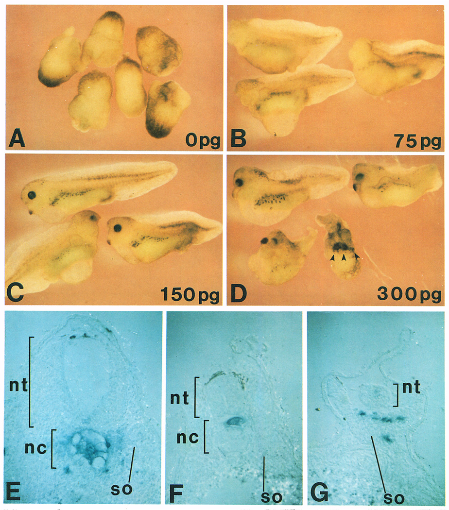
A Xenopus gene whose expression can be activated by the organizer-specific homeobox genes goosecoid and Xnot2 was isolated by differential screening. The chordin gene encodes a novel protein of 941 amino acids that has a signal sequence and four Cys-rich domains. The expression of chordin starts in Spemann's organizer subsequent to that of goosecoid, and its induction by activin requires de novo protein synthesis. Microinjection of chordin mRNA induces twinned axes and can completely rescue axial development in ventralized embryos. This molecule is a potent dorsalizing factor that is expressed at the right time and in the right place to regulate cell-cell interactions in the organizing centers of head, trunk, and tail development.


Figures

Figure 1
Figure 1. chordin Is Expressed in Regions with Head, Trunk, and Tail Organizer Activity
Digoxygenin-labeled antisense chordin RNA was hybridized to embryos at stage 9.5 (A); stage 10.25 (B); stage 10.75 (C); stage 11.5 (D); stage 13, note expression in the prechordal plate and notochord (E); stage 26 (F); stage 33, note expression in the chordoneural hinge of the tailbud (G); stage 33, enlarged view of the tailbud region (G’); stage 42, enlarged view of the tail region (H); LiCl-treated embryo at stage 11.5 (I), compare with (D). (A)–(D) and (I) are vegetal views, dorsal side is at the top. (E) is viewed from the dorsal side with anterior at top. (F)–(H) are lateral views.
Figure 2
Figure 2. chordin, but Not noggin, Expression Is Activated by gsc and Xnot2 Homeobox-Containing mRNAs
Embryos were injected in the equator with the indicated synthetic mRNA at the 8-cell stage, cultured until stage 11, and hybridized with a chordin (A–F) or noggin (G–I) antisense RNA probe. Four radial injections were given to wild-type embryos, and two diagonal ones to UV-treated embryos. (A) Wild-type (wt) uninjected embryo hybridized with the chordin probe. Staining is only seen in the organizer region. (B) Sibling embryo radially injected with gsc mRNA (80 pg per blastomere). Arrows indicate ectopic expression of chordin. (C) UV-treated embryo, chordin expression is abolished. (D) UV-treated embryo injected diagonally at two sites with gsc mRNA, arrows indicate two spots in which chordin expression is activated. (E) Embryo injected radially with Xnot2 mRNA (100 pg per blastomere). Arrows indicate regions of ectopic chordin expression. (F) Embryo injected radially with a biologically active homeobox mRNA of the Antennapedia type (XIHbox-1); no ectopic expression of chordin was detected even at 500 pg per blastomere. (G) Wild-type embryo hybridized with the noggin probe. Because the noggin signal is deep, embryos were cleared in Murray’s solution. (H) Embryo treated as in (G), but radially injected with gsc mRNA. Note that noggin is not activated by gsc. (I) Embryo treated as (G), but radially injected with Xnot2 mRNA, there is no ectopic expression of noggin. None of the RNAs injected in this figure cross-hybridized with the probes used, as determined in control experiments. Photos were taken from the vegetal side.
Figure 3
Figure 3. chordin Induction by Activin Requires De Novo Protein Synthesis
(A) Temporal pattern of chordin expression. Northern blot analysis was performed with total RNA (7.5 µg) from various stages of early Xenopus embryos. Full-length chordin (chd) or gsc (gsc) cDNA were used as probes. 18S RNA stained with ethidium bromide is shown below as a loading control. The chordin transcript was first detected at stage 9.5, 1 hr before the onset of gastrulation. The accumulation of zygotic gsc RNA was detected earlier, at stage 9, 2 hr before gastrulation, as previously described (Cho et al., 1991). Maternal transcripts (E, egg) are present in the case of gsc but not in that of chordin, even after longer exposure. (B) CHX inhibits activation of the chordin gene by activin. Animal caps (stage 8) were treated with 30 ng/ml activin for 2.5 hr (corresponding to stage 10) in the presence or absence of a protein synthesis inhibitor CHX (5 µg/ml) (Rosa, 1989; Cho et al., 1991). Total RNA (10 µg) was loaded in this Northern blot. Lane 1, untreated control; lane 2, CHX alone; lane 3, activin alone; lane 4, activin in the presence of CHX. Note that while chordin (chd) induction is inhibited by blocking protein synthesis, the induction of gsc and of noggin (nog) is somewhat increased. This indicates that while noggin and gsc are primary response genes to activin treatment, chordin is a secondary response gene.
Figure 4
Figure 4. Xenopus chordin Encodes a Putative Secreted Protein
(A) The amino acid sequence deduced from the nucleotide sequence of a full-length Xenopus chordin cDNA clone is shown. The initiator Met was assigned to the first ATG in the longest reading frame, which has an in-frame stop codon 130 bp upstream of this ATG. Four potential N-glycosylation consensus sequences are indicated by asterisks. Four internal repeats are underlined. A hydrophobic signal peptide segment is found at the amino terminus (residues 1–19). (B) Schematic structure of chordin protein. The potential signal peptide and four internal Cys-rich repeats are shown by closed and open boxes, respectively. Vertical bars indicate potential N-glycosylation sites. SP, signal peptide. (C) Comparison of Cys-rich repeats in chordin and those in some secreted proteins. The residues conserved among four repeats are boxed. chd, Xenopus chordin; TSP-2, mouse thombospondin-2; α-PC, human α1 procollagen type I; vWBF, human von Willebrand factor C1 domain.
Figure 5
Figure 5. chordin mRNA Induces Secondary Axes
(A) Secondary axis formed after microinjection of chordin RNA (200 pg) into a ventrovegetal blastomere of an 8-cell embryo. (A’) Immunostaining with a notochord marker (MZ-15) of the same embryo shown in (A). A second notochord (II) and extra auditory vesicles (arrowheads) are present. (B) Secondary tail formed after microinjection of chordin RNA into the animal pole. (B’) Immunostaining with MZ-15 notochord antibody. No mature second notochord was found in this secondary tail. C) A dorsalized embryo resulting from the injection of chordin RNA (200 pg) into a dorsovegetal blastomere of an 8-cell embryo. Notochord staining showed a short and thick double-barreled notochord (data not shown). D) A double-headed embryo induced by chordin. chordin RNA (150 pg) was coinjected with β-galactosidase RNA into a A4 blastomere of a 32-cell embryo. Cement glands (arrowheads) and eyes (arrows) are duplicated. Strong β-galactosidase activity was detected in the posterior epidermis but not in the secondary axis (confirmed by histological section; data not shown), indicating that chordin has noncell-autonomous effects on uninjected cells. In all cases, injection of β-galactosidase or prolactin control RNAs showed no particular phenotypes.
Figure 6
Figure 6. Dose-Dependent Axial Rescue by chordin mRNA
Increasing doses of chordin mRNA were injected into one vegetal blastomere of an 8-cell embryo after UV irradiation at the 1-cell stage. (A) Uninjected embryos that have been completely lost axial structures after UV treatment (DAI = 0.1, n = 10); (B) 75 pg of chordin RNA (DAI = 2.1, n = 17); (C) 150 pg of chordin RNA (DAI = 4.2, n = 10); (D) 300 pg of chordin RNA (DAI = 6.2, n = 8). Multiple cement glands are indicated by arrowheads. Injection of control prolactin RNA did not rescue the UV-ventralized phenotype. (E–G) transverse histological sections of embryos injected with β-galactosidase and chordin mRNA into the C-tier region and stained with X-Gal. Note that the injected cells populate in the dorsal axis and recruit uninjected cells into the axis. nt, neural tube; nc, notochord; so, somite.

References

    1. Amaya E, Musci TJ, Kirschner MW. Expression of a dominant negative mutant of the FGF receptor disrupts mesoderm formation in Xenopus embryos. Cell. 1991;66:257–270. - PubMed
    1. Blumberg B, Wright CVE, De Robertis EM, Cho KWY. Organizer-specific homeogenes in Xenopus laevis embryos. Science. 1991;253:194–196. - PubMed
    1. Bornstein P. Thrombospondins: structure and regulation of expression. FASEB J. 1992;6:3290–3299. - PubMed
    1. Cho KWY, Blumberg B, Steinbeisser H, De Robertis EM. Molecular nature of Spemann’s organizer: the role of the Xenopus homeobox gene goosecoid. Cell. 1991;67:1111–1120. - PMC - PubMed
    1. Christian JL, Moon RT. Interactions between Xwnt-8 and Spemann organizer signaling pathways generate dorsoventral pattern in the embryonic mesoderm of Xenopus. Genes Dev. 1993;7:13–28. - PubMed

Publication types

MeSH terms

Associated data