. 1994 Jul;135(1):365-72.
doi: 10.1210/endo.135.1.8013370.

Voltage-independent calcium channels mediate slow oscillations of cytosolic calcium that are glucose dependent in pancreatic beta-cells

Affiliations

Voltage-independent calcium channels mediate slow oscillations of cytosolic calcium that are glucose dependent in pancreatic beta-cells

C A Leech et al. Endocrinology. 1994 Jul.

Abstract

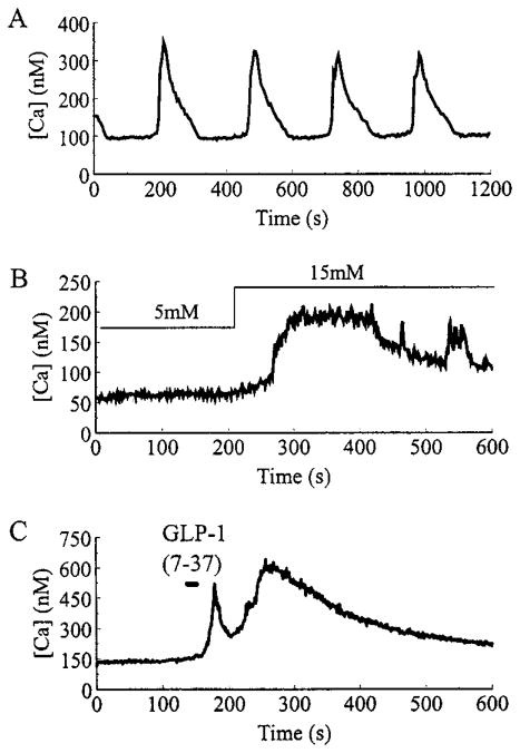
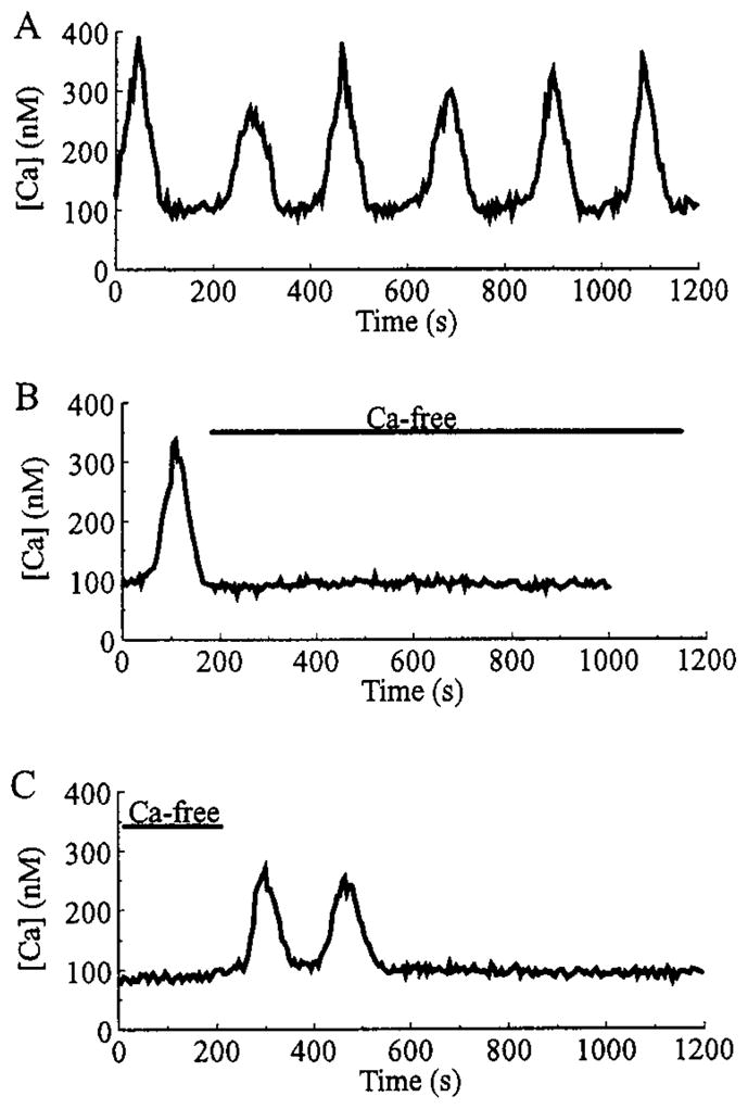
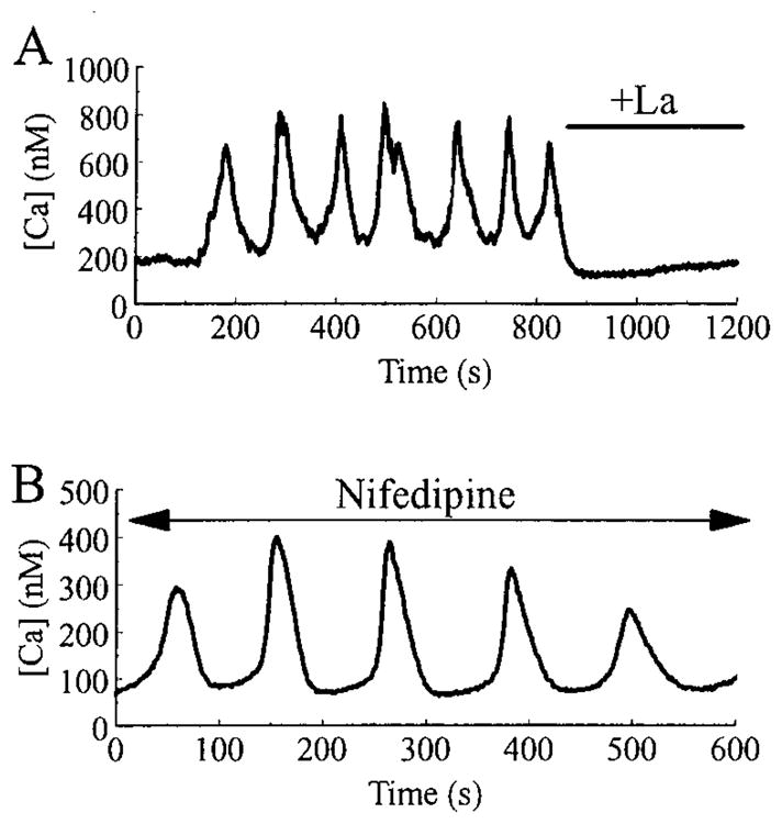
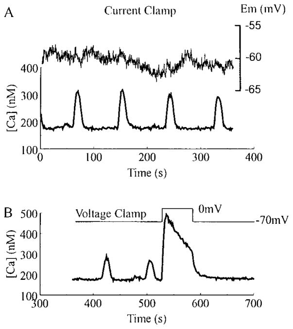
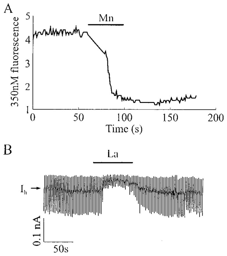
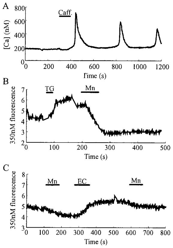
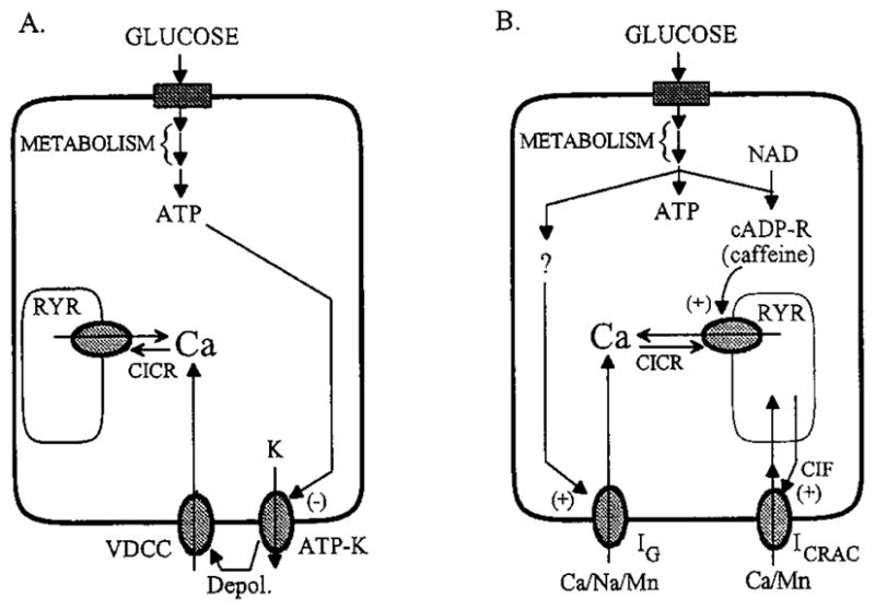
Pancreatic beta-cells and HIT-T15 cells exhibit oscillations of cytosolic calcium ([Ca2+]i) that are dependent on glucose metabolism and appear to trigger pulsatile insulin secretion. Significantly, differences in the pattern of this [Ca2+]i oscillatory activity may have important implications for our understanding of how glucose homeostasis is achieved during the feeding and fasting states. When single beta-cells are exposed to a stepwise increase in glucose concentration that mimics the transition from fasting to feeding states, fast irregular oscillations of [Ca2+]i are observed. Alternatively, when single beta-cells are equilibrated in a steady state concentration of glucose that mimics the fasting state, slow periodic oscillations of [Ca2+]i are noted. Here we report a fundamental difference in the mechanism by which glucose induces these two types of [Ca2+]i oscillatory activity. In agreement with previous studies, we substantiate a role for L-type voltage-dependent Ca2+ channels as mediators of the fast oscillations of [Ca2+]i observed after a stepwise increase in glucose concentration. In marked contrast, we report that voltage-independent calcium channels (VICCs) mediate slow oscillations of [Ca2+]i that occur when beta-cells are equilibrated in steady state concentrations of glucose. Slow [Ca2+]i oscillations are mediated by VICCs which are pharmacologically and biophysically distinguishable from voltage-dependent Ca2+ channels that mediate fast oscillations. Specifically, slow [Ca2+]i oscillations are blocked by extracellular La3+, but not by nifedipine, and are independent of changes in membrane potential. Measurement of membrane conductance also indicate an important role for VICCs, as demonstrated by a steady state inward Ca2+ current that is blocked by La3+. The steady state Ca2+ current appears to generate slow [Ca2+]i oscillations by triggering Ca(2+)-induced Ca2+ release from intracellular Ca2+ stores, a process that is mimicked by extracellular application of caffeine, a sensitizer of the ryanodine receptor/Ca2+ release channel. Depletion of intracellular Ca2+ stores with thapsigargin stimulated Mn2+ influx, suggesting the presence of Ca(2+)-release-activated Ca2+ channels. Taken together, these observations are consistent with a role for VICCs (possibly G-type channels) and/or Ca(2+)-release-activated Ca2+ channels as mediators of slow [Ca2+]i oscillations in beta-cells. We propose that slow oscillations of [Ca2+]i probably serve as important initiators of insulin secretion under conditions in which tight control of glucose homeostasis is necessary, as is the case during the fasting normoglycemic state.


Figures

Fig. 1
Fig. 1
Representative examples of glucose and insulinotropic peptide-induced alterations of [Ca]i in β-cells. A, Slow oscillations of [Ca2+]i recorded when a β-cell was equilibrated in a steady state concentration (7.5 mM) of glucose. B, A β-cell was initially equilibrated in 5 mM glucose, then a stepwise increase in glucose concentration to 15 mM was applied by rapidly switching the contents of the bathing solution. C, A 30-sec pulse of 10 nM GLP-1-(7-37) was directly applied to a cell bathed in 5 mM glucose, using a puffer pipette (see Materials and Methods).
Fig. 2
Fig. 2
Slow oscillations of [Ca2+]i require extracellular Ca2+. A, A β-cell exhibited slow oscillations of [Ca2+]i when the bath solution contained a normal concentration (2.6 mM) of Ca2+. As illustrated in B, when the superfusate was switched to a nominally Ca2+-free saline (as indicated by the bar), the oscillations were abolished. As illustrated in C, reintroduction of 2.6 mM Ca2+ briefly restored the slow oscillatory activity. All recordings are from the same cell under conditions of steady state equilibration in 7.5 mM glucose. Records A and B are separated by a 350-sec interval, whereas records B and C are separated by a 200-sec interval.
Fig. 3
Fig. 3
Slow oscillations of [Ca2+]i are blocked by La3+, but not by nifedipine. In A, slow [Ca2+]i oscillations were recorded from a HIT-T15 cell bathed in a solution containing 2 mM glucose and 2.6 mM Ca2+. Note that the oscillations were rapidly blocked by superfusion with an identical saline solution to which 500 μM La3+ was added. In B, slow [Ca2+]i oscillations recorded from a HIT-T15 cell bathed in 2 mM glucose and 2.6 mM Ca2+ were unaffected by the addition of 5 μM nifedipine to the superfusate.
Fig. 4
Fig. 4
Slow oscillations of [Ca2+]i are independent of changes in membrane potential. In A and B are illustrated perforated patch recordings from the same fura-2AM-loaded HIT-T15 cell equilibrated in 2 mM glucose. As indicated in A, [Ca2+]i (lower trace, left side scale) and the resting membrane potential (Em; upper trace, right side scale) were simultaneously monitored in the current clamp recording configuration. Little or no change in membrane potential was observed during the [Ca2+]i oscillations. As illustrated in B, the cell was then voltage clamped to a holding potential of −70 mV. At this holding potential, the slow oscillations were not blocked (note different scales for [Ca2+]i in A and B). Application of a depolarizing voltage step to 0 mV induced a large rise in [Ca2+]i due to the opening of VDCCs, thereby confirming that electrical access to the cell was intact. Similar results were obtained with rat β-cells.
Fig. 5
Fig. 5
HIT-T15 cells equilibrated in 2 mM glucose exhibit a pathway for divalent cation influx that is permeant to Mn2+, but blocked by La3+. In A is illustrated quenching of the intracellular fura-2 fluorescence signal by 2.6 mM Mn2+ applied extracellularly from a puffer pipette (Ca2+ was omitted from the Mn2+-containing solution). Fura-2 was excited at 350 nm, and the fluorescence emmision was measured at 510 nm. In B is illustrated a voltage-clamp experiment in which the resting membrane conductance was monitored by applying ±10 mV voltage steps (each for 1 sec) from a holding potential of −80 mV. La3+ (100 μM), applied extracellularly from a puffer pipette, caused a reduction in membrane conductance (as indicated by the smaller current responses to applied voltage steps) and a shift in the holding current (IH) in the outward direction.
Fig. 6
Fig. 6
Evidence for a ICRAC. In A, a rat β-cell, equilibrated in 7.5 mM glucose, responded to a single 60-sec application of caffeine (caff.; 10 mM) with a series of slow oscillations of [Ca2+]i. In B, a HIT-T15 cell equilibrated in 2 mM glucose was stimulated with 2 μM thapsigargin (TG), causing a rise in [Ca2+]i, detected as a small increase in the intensity of fluorescence due to excitation of fura-2 at 350 nm. A subsequent application of 2.6 mM Mn2+ (substituted for Ca2+) caused quenching of the fluorescence signal in six of seven cells, indicating activation of a Mn2+-permeant conductance by thapsigargin. No quenching by Mn2+ was observed before application of thapsigargin. In C, a HIT-T15 cell equilibrated in 2 mM glucose was treated with 2.6 mM Mn2+ before and after stimulation with 10 μM econazole (EC). Mn2+-induced quenching of the fura-2 fluorescence was not increased after treatment with econazole (n = 7). Note that econazole caused a rise in [Ca2+]i, measurable as an increase in the intensity of fura-2 fluorescence.
Fig. 7
Fig. 7
A, Glucose-signaling pathways that may mediate the rise in [Ca2+]i observed after a stepwise increase in glucose concentration. The uptake and metabolism of glucose generate ATP, which acts to depolarize (depol.) β-cells by inhibiting ATP-sensitive potassium channels (ATP-K) that regulate the resting membrane potential. Membrane depolarization leads to the activation of VDCC. Influx of Ca2+ through these channels causes the elevation of cytosolic [Ca2+], which also triggers CICR through activation of the ryanodine receptor/calcium release channels (RYR). B, For a cell equilibrated in a fixed concentration of glucose below the threshold for initiation of electrical bursting, metabolism of glucose may generate multiple signals. These are proposed to include ATP, cyclic ADP-ribose (cADP-R) generated from nicotinamide adenine dinucleotide (NAD), and an as yet to be characterized metabolite (?) that activates the G-type channel (IG). Influx of Ca2+ through the G-channel raises [Ca2+]i until it reaches a level that stimulates CICR. Sensitization of the ryanodine receptors to Ca2+ may result from the action of cADP-R on them (37). Release of Ca2+ from intracellular stores and subsequent depletion of the stores leads to activation of a ICRAC by a CIF or a small mol wt G-protein (23). Further elevation of [Ca2+]i then inhibits release from the stores, and a combination of Ca2+ extrusion from the cell and reuptake by the stores brings [Ca2+]i back to basal levels.

References

    1. Holz GG, IV, Kuhtreiber WM, Habener JF. Pancreatic β-cells are rendered glucose competent by the insulinotropic hormone GLP-1(7–37) Nature. 1993;361:362–365. - PMC - PubMed
    1. Corkey BE, Deeney JT, Glennon MC, Matschinsky FM, Prentki M. Regulation of steady-state free Ca2+ levels by the ATP/ADP ratio and orthophosphate in permeabilized RINm5F cells. J Biol Chem. 1988;263:4247–4253. - PubMed
    1. Woods NM, Cuthbertson KSR, Cobbold PH. Repetitive transient rises in cytoplasmic free calcium in hormone-stimulated hepatocytes. Nature. 1986;319:600–602. - PubMed
    1. Berridge MJ. Inositol trisphosphate and calcium signalling. Nature. 1993;361:315–325. - PubMed
    1. Schlegal W, Winiger BP, Mollard P, Vacher P, Wuarin F, Zahnd GR, Wollheim CB, Dufy B. Oscillations of cytosolic Ca2+ in pituitary cells due to action potentials. Nature. 1987;329:719–721. - PubMed

Publication types