

Labeling of cerebral amyloid in vivo with a monoclonal antibody
- PMID: 8021711
- PMCID: PMC9887729
- DOI: 10.1097/00005072-199407000-00009
Labeling of cerebral amyloid in vivo with a monoclonal antibody
Abstract
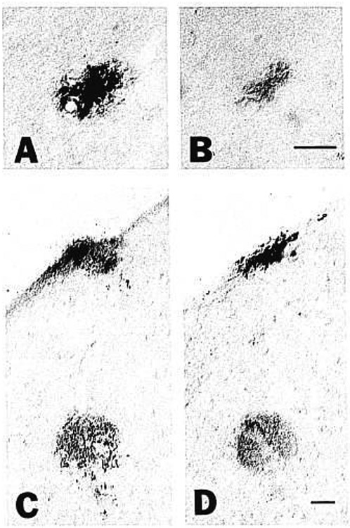
We assessed the ability of a murine monoclonal antibody to bind selectively to beta-amyloid in the brains of living nonhuman primates. To circumvent the blood-brain barrier, we injected unlabeled antibody 10D5 (murine whole IgG1 and/or Fab fragments) into the cerebrospinal fluid of the cisterna magna in three aged monkeys. A control animal was given an intracisternal injection of nonimmune mouse whole IgG plus Fab. Twenty-four hours later, the animals were perfused and prepared for immunohistochemical detection of bound murine immunoglobulin in brain. All three experimental animals showed selective binding of 10D5 to approximately 5-15% of amyloid deposits in cerebral cortex, primarily near the cortical surface. There was no labeling in the control animal. In vivo-labeled deposits were confirmed to be beta-amyloid by electron microscopy and by in vitro immunohistochemistry in adjacent sections. The animals tolerated the injection well, although some polymorphonuclear leukocytes infiltrated portions of the subarachnoid space and superficial neocortex. These results provide the first demonstration that it may be feasible to selectively direct a tagged monoclonal antibody to beta-amyloid in the brain for therapeutic or diagnostic purposes. With enhancement of labeling efficiency, the method also may be useful for studying the progression of beta-amyloidosis in experimental animals using emission tomography.
Figures


Similar articles
-
Animal models of cerebral beta-amyloid angiopathy.Brain Res Brain Res Rev. 1997 Sep 30;25(1):70-84. doi: 10.1016/s0165-0173(97)00017-9. Brain Res Brain Res Rev. 1997. PMID: 9370051 Review.
-
Non-Fc-mediated mechanisms are involved in clearance of amyloid-beta in vivo by immunotherapy.J Neurosci. 2002 Sep 15;22(18):7873-8. doi: 10.1523/JNEUROSCI.22-18-07873.2002. J Neurosci. 2002. PMID: 12223540 Free PMC article.
-
Circulating amyloid-beta peptide crosses the blood-brain barrier in aged monkeys and contributes to Alzheimer's disease lesions.Vascul Pharmacol. 2002 Jun;38(6):303-13. doi: 10.1016/s1537-1891(02)00198-2. Vascul Pharmacol. 2002. PMID: 12529925
-
Fusion of hIgG1-Fc to 111In-anti-amyloid single domain antibody fragment VHH-pa2H prolongs blood residential time in APP/PS1 mice but does not increase brain uptake.Nucl Med Biol. 2015 Aug;42(8):695-702. doi: 10.1016/j.nucmedbio.2015.03.003. Epub 2015 Mar 18. Nucl Med Biol. 2015. PMID: 25960433
-
Distribution of beta/A4 protein and amyloid precursor protein in hereditary cerebral hemorrhage with amyloidosis-Dutch type and Alzheimer's disease.Am J Pathol. 1993 May;142(5):1449-57. Am J Pathol. 1993. PMID: 7684195 Free PMC article. Review.
Cited by
-
Vector-mediated delivery of 125I-labeled beta-amyloid peptide A beta 1-40 through the blood-brain barrier and binding to Alzheimer disease amyloid of the A beta 1-40/vector complex.Proc Natl Acad Sci U S A. 1995 Oct 24;92(22):10227-31. doi: 10.1073/pnas.92.22.10227. Proc Natl Acad Sci U S A. 1995. PMID: 7479757 Free PMC article.
-
Insulysin hydrolyzes amyloid beta peptides to products that are neither neurotoxic nor deposit on amyloid plaques.J Neurosci. 2000 Dec 1;20(23):8745-9. doi: 10.1523/JNEUROSCI.20-23-08745.2000. J Neurosci. 2000. PMID: 11102481 Free PMC article.
-
Drug targeting of a peptide radiopharmaceutical through the primate blood-brain barrier in vivo with a monoclonal antibody to the human insulin receptor.J Clin Invest. 1997 Oct 1;100(7):1804-12. doi: 10.1172/JCI119708. J Clin Invest. 1997. PMID: 9312181 Free PMC article.
-
The cerebral proteopathies: neurodegenerative disorders of protein conformation and assembly.Mol Neurobiol. 2000 Feb-Apr;21(1-2):83-95. doi: 10.1385/MN:21:1-2:083. Mol Neurobiol. 2000. PMID: 11327151 Review.
-
The ART of loss: Abeta imaging in the evaluation of Alzheimer's disease and other dementias.Mol Neurobiol. 2008 Aug;38(1):1-15. doi: 10.1007/s12035-008-8019-y. Epub 2008 Aug 9. Mol Neurobiol. 2008. PMID: 18690556 Review.
References
-
- Glenner GG, Wong CW. Alzheimer's disease: Initial report of the purification and characterization of a novel cerebrovascular amyloid protein. Biochem Biophys Res Commun 1984; 120:885–890 - PubMed
-
- Kemper T. Neuroanatomical and neuropathological changes in normal aging and in dementia. In: Albert ML, ed. Clinical Neurology of Aging. New York: Oxford University Press, 1984: 9–52
-
- Mandybur TI. Cerebral amyloid angiopathy: The vascular pathology and complications. J Neuropathol Exp Neurol 1986; 45:79–90 - PubMed
Publication types
MeSH terms
Substances
Grants and funding
LinkOut - more resources
Full Text Sources
Other Literature Sources
