

Locus-specific analysis of human leukocyte antigen class I expression in melanoma cell lines
- PMID: 8081556
- PMCID: PMC2561308
- DOI: 10.1097/00002371-199407000-00002
Locus-specific analysis of human leukocyte antigen class I expression in melanoma cell lines
Abstract
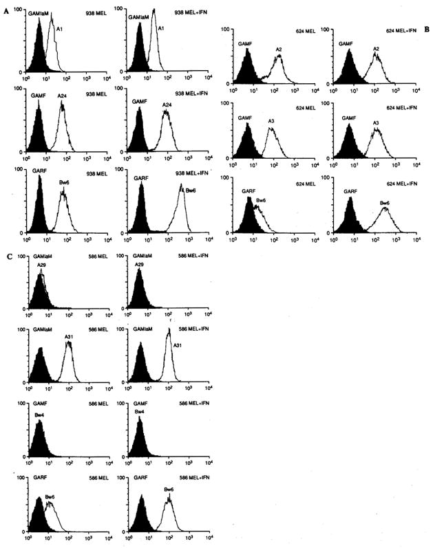
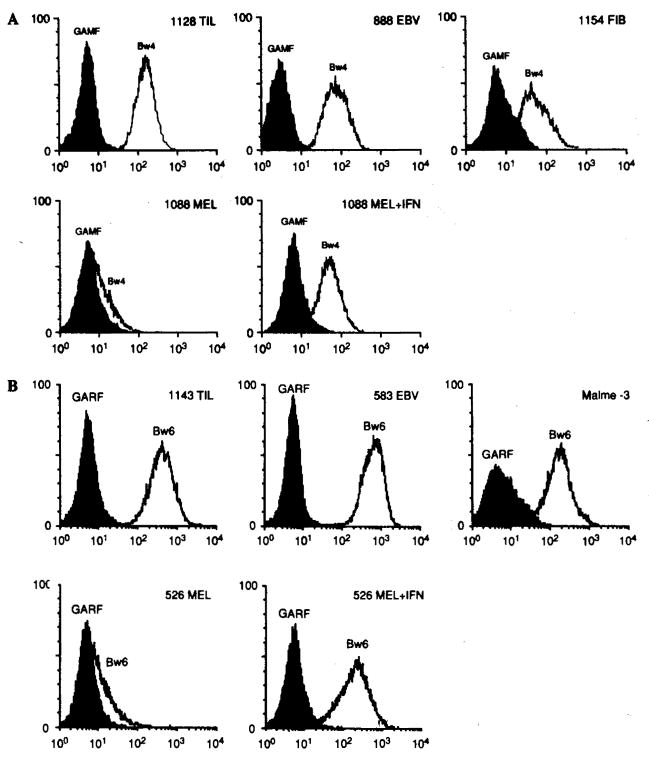
Surface expression of human leukocyte antigen (HLA) class I antigens on melanoma lines was evaluated by locus-specific monoclonal antibodies (mAbs) with three different techniques: Fluorescence-activated cell sorting (FACS), immunohistochemistry with cytospin preparation (ICP), and complement-mediated cytotoxicity (CMC). Eleven HLA class I-expressing cell lines developed from metastases were used. Specific expression of HLA loci was examined under routine culture conditions and after 48-h incubation in interferon-gamma (IFN-gamma; 500 U/ml). Loss of allelic expression was seen in one line (586-MEL): Products of genes coding for HLA-A29 and -B44, in strong linkage disequilibrium, were not detectable. HLA-A antigens were consistently detected by all methodologies and minimally affected by pretreatment with IFN-gamma. HLA-B antigens were detectable in 8 of 11 lines by ICP and 3 of 11 lines by CMC. By FACS the supratypic specificity HLA-Bw6 was expressed at low levels in most lines (mean fluorescence 47.2 +/- 13.4 and rose to 259.8 +/- 45.9 after incubation with IFN-gamma; p < 0.001). HLA-Cw antigen detection by CMC correlated with HLA-B (p < 0.01), suggesting that down-regulation and sensitivity to IFN-gamma are shared by the two loci. This low expression of the HLA-B antigens may play a role in the evasion of the host immune response and its up-regulation may be useful in allowing tumor antigen recognition.
Figures
References
-
- Weber JS, Rosenberg SA. Effects of murine tumor class I major histocompatibility complex expression on antitumor activity of tumor-infiltrating lymphocytes. J Nail Cancer Inst. 1990;82:755–61. - PubMed
-
- Crowley NJ, Darrow TL, Quinn-Allen MA, Seigler HF. MHC-restricted recognition of autologous melanoma by tumor-specific cytotoxic T cells. Evidence for restriction by a dominant HLA-A allele. J Immunol. 1991;146:1692–9. - PubMed
-
- Horn SS, Topalian SL, Simonis T, Mancini M, Rosenberg SA. Common expression of melanoma tumor associated antigens recognized by human tumor infiltrating lymphocytes: analysis by human lymphocyte antigen restriction. J Immunother. 1991;10:153–64. - PubMed
MeSH terms
Substances
Grants and funding
LinkOut - more resources
Full Text Sources
Other Literature Sources
Medical
Research Materials