Comparative Study
. 1994 Apr 15;2(4):271-82.
doi: 10.1016/s0969-2126(00)00029-0.

Functional implications of quasi-equivalence in a T = 3 icosahedral animal virus established by cryo-electron microscopy and X-ray crystallography

Affiliations
Comparative Study

Functional implications of quasi-equivalence in a T = 3 icosahedral animal virus established by cryo-electron microscopy and X-ray crystallography

R H Cheng et al. Structure. .

Abstract

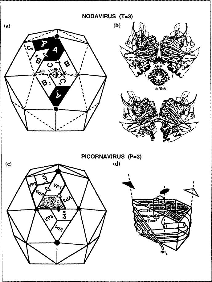
Background: Studies of simple RNA animal viruses show that cell attachment, particle destabilization and cell entry are complex processes requiring a level of capsid sophistication that is difficult to achieve with a shell containing only a single gene product. Nodaviruses [such as Flock House virus (FHV)] are an exception. We have previously determined the structure of FHV at 3 A resolution, and now combine this information with data from cryo-electron microscopy in an attempt to clarify the process by which nodaviruses infect animal cells.

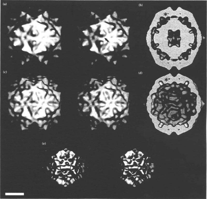
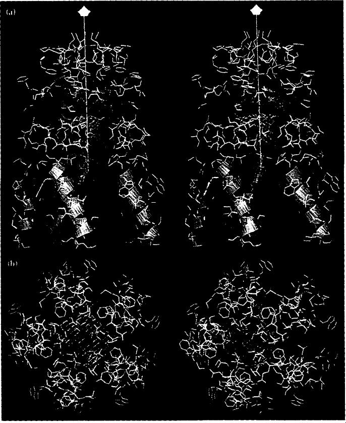
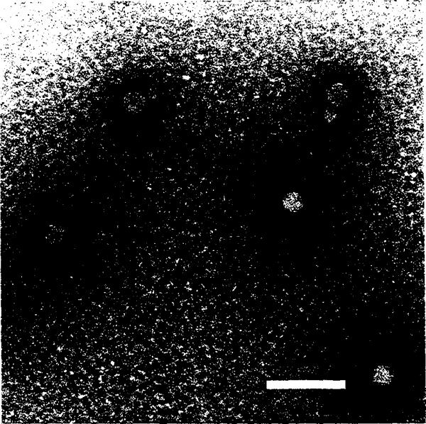
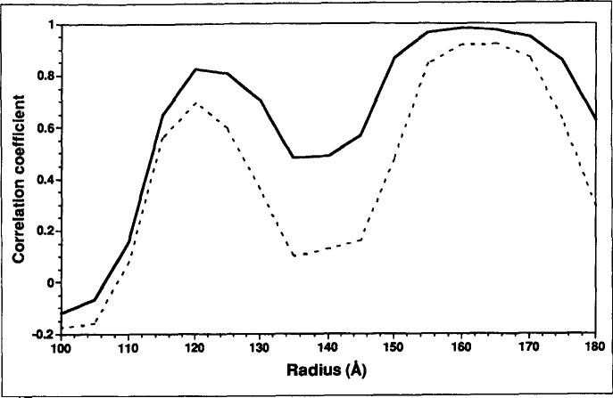
Results: A difference map was computed in which electron density at 22 A resolution, derived from the 3.0 A resolution X-ray model of the FHV capsid protein, was subtracted from the electron density derived from the cryo-electron microscopy reconstruction of FHV at 22 A resolution. Comparisons of this density with the X-ray model showed that quasi-equivalent regions of identical polypeptide sequences have markedly different interactions with the bulk RNA density. Previously reported biphasic kinetics of particle maturation and the requirement of subunit cleavage for particle infectivity are consistent with these results.

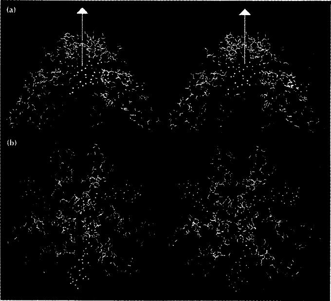
Conclusions: On the basis of this study we propose a model for nodavirus infection that is conceptually similar to that proposed for poliovirus but differs from it in detail. The constraints of a single protein type in the capsid lead to a noteworthy use of quasi-symmetry not only to control the binding of a 'pocket factor' but also to modulate maturation cleavage and to release a pentameric helical bundle (with genomic RNA attached) that may further interact with the cell membrane.


Figures

Fig. 1
Fig. 1
A comparison of a T=3 nodavirus capsid with a P=3 picornavirus capsid, showing that the two capsids are similar in overall shape, size and organization. (a) Nodavirus capsid. (b) Side views of the C–C 2 contact (top) and A–Bs contact (bottom). (c) Picornavirus capsid. (d) Schematic representation of the eight-stranded β-barrel structure of the capsid subunits. Closed symbols identify icosahedral axes of symmetry whereas open symbols identify quasi (lo-cal) symmetry axes.
Fig. 2
Fig. 2
Stereoview of the γ-peptides and ordered RNA segments of the FHV particle structure. The triangle represents the border of the icosahedral asymmetric unit containing A, B and C in the T= 3 particle in Fig. 1. Blue helices are associated with the A subunits (γA), red with the B subunits (γB), and green with the C subunits (γc). Quasi six-fold axes are shown as red triangles, the five-fold axes as a red pentagon. Duplex RNA is represented as ball-and-stick models.
Fig. 3
Fig. 3
Frozen-hydrated Flock House virus. Polyoma virus (larger particles, 495 Å diameter) was mixed with the FHV sample prior to the vitrification for size comparison and as a familiar specimen which aided assessment of image quality. Bar = 1000Å.
Fig. 4
Fig. 4
Electron density distributions displayed as surface shaded images of FHV. Bar = 100 Å (a) Stereoview of FHV from the cryoEM reconstruction. The arrow indicates the direction of view in Fig. 5. (b) Equatorial section of (a), showing the interior RNA density. The large center cavity may be an artificial feature because the noise in image reconstruction of icosahedral particles is greatest in this region. One of the 16 A cavities is labeled with an asterisk. Four of the 60 cavities are visible. (c) Stereoview of the 22 Å electron density map, computed with structure factors based on the 3.0 Å resolution model [13]. (d) Equatorial section of (c). Note the 16A cavity (asterisk) and the absence of interior RNA density. (e) Stereoview derived from the cryoEM -X-ray model difference map. This electron density not accounted for by the X-ray model is largely RNA.
Fig. 5
Fig. 5
Close up of the two-fold related protrusions at the quasi threefold axes of the surface (arrowed in Fig. 4a) showing the consistency of the cryoEM and X-ray data. (a) CryoEM electron density (blue contours) superimposed on the FHV X-ray model (yellow and green ribbon diagram). (b) Cross-section view of the full virion (blue). Superimposed on this are; FHV protein Cα atomic coordinates (yellow), the contoured difference electron density map (red lines), and the locally ordered RNA (yellow, red and blue space-filling representation). Vertical white line represents icosahedral two-fold axis; other white lines capped with pentagon symbols identify two five-fold axes.
Fig. 6
Fig. 6
Positions of the γ-helices. (a) Cut away view of the whole FHV virion. Electron density derived from the cryoEM reconstruction is blue, density derived from the difference map is red. γy8-helices are red cylinders, γc-helices green and γA-helices light blue. Asterisks indicate the helices shown in close up in (b). (b) Close up of γB and γc and two-fold related mates showing the close association with bulk RNA (red contours) and ordered RNA (red, blue and yellow space-filling model). (c) Side view of a bundle of γA-helices. The five-fold axis (vertical white line with red pentagon) passes through the ellipsoidal density (red contours indicate the density as plotted in the difference map, white contours the density from the X-ray map and blue contours the cryoEM envelope). One A subunit is shown in ribbon representation (green, yellow and red), others as thin yellow lines (Coc backbone trace).
Fig. 7
Fig. 7
Stereoviews of the helical bundle of γ-peptides at the pentamer contact and residues from the subunits adjacent to the five-fold axis (labeled A in Fig. 1a). (a) Side view (five-fold axis is shown as a white line with a pentagon symbol at the end furthest from the virus center). (b) View directly down the five-fold axis. Residues in yellow are hydrophobic and those in red are charged. Blue helices are the γA-peptides. Blue contours are the ellipsoidal density seen in the X-ray map, red contours correspond to this density from the difference map.
Fig. 8
Fig. 8
Stereoviews of the γB- and γc-peptides (red and green helices, respectively) and the peptide arms (green, space-filling atoms) at the quasi six-fold contact and residues from the subunits adjacent to the quasi six-fold axis (labeled B and C in Fig. 1a). (a) Side view with the quasi six-fold axis shown as a white line with a triangle at the end. (b) A view directly down the quasi sixfold axis.
Fig. 9
Fig. 9
Micrograph of negatively-stained FHV particles that were heated to 65°C. Most of the particles exhibit a 'puff of density that appears to protrude from a single site on the capsid. The novel structure seen at each of the FHV pentamer axes makes it a strong candidate for the location of release of a y-helical bundle and associated RNA. Bar = 1000 Å.
Fig. 10
Fig. 10
Plot of correlation coefficients as a function of virus radius, comparing the X-ray model with cryoEM reconstructions before (dashed line) and after (solid line) corrections were made to reduce the effects of the microscope contrast transfer function. The plots clearly demonstrate marked improvement in the data within the capsid shell after compensation for the defocus effects were included.

References

    1. Caspar DLD, Klug A. Physical principles in the construction of regular viruses. Cold Spring Harbor Symp. Quant. Biol. 1962;27:1–24. - PubMed
    1. Johnson JE, Fisher AJ. Principles of virus structure. In: Webster RG, Granoff A, editors. Encyclopedia of Virology. Academic Press; London: 1994. in press.
    1. Rossmann MG, Johnson JE. Icosahedral RNA virus structure. Annu. Retr Biochem. 1989;58:533–573. - PubMed
    1. Chen Z, et al. Johnson JE. Protein-RNA interactions in an icosahedral virus at 3.OÅ resolution. Science. 1989;245:154–159. - PubMed
    1. Olson NH, et al. Rossmann MG. Structure of a human rhinovirus complexed with its receptor molecule. Proc. Natl. Acad Sci. USA. 1993;90:507–511. - PMC - PubMed

Publication types