. 1993 Feb 15;190(3):724-31.
doi: 10.1006/bbrc.1993.1109.

Expression of extracellular ligand-binding domain of murine guanylate cyclase/atrial natriuretic factor receptor cDNA in Escherichia coli

Affiliations

Expression of extracellular ligand-binding domain of murine guanylate cyclase/atrial natriuretic factor receptor cDNA in Escherichia coli

K N Pandey et al. Biochem Biophys Res Commun. .

Abstract

The membrane-bound form of guanylate cyclase/atrial natriuretic factor receptor (GC/ANF-R) is a 135 kDa transmembrane glycoprotein which binds ANF with high affinity. We have expressed the extracellular ligand-binding domain of murine guanylate cyclase ANF-R (GC/ANFR-LBD) cDNA in Escherichia coli. The cDNA encoding the extracellular ANF-binding domain (nucleotide positions covering from 432-1755 base pair) of GC/ANF-R was amplified by polymerase chain reaction, cloned into BamHI site of pGEX-3X prokaryotic expression vector and was transfected into E. coli, strain JM101. After isopropyl-beta-D-thiogalactopyranoside (IPTG) induction of bacterial cells, the GC/ANFR-LBD was expressed as the glutathione-S-transferase (GST) fusion protein, yielding a molecular mass of 70 kDa. The expressed fusion protein was characterized for binding affinity to both full length and truncated ANF molecules. After expression in E. coli, the binding of 125I-ANF to the extracellular region of GC/ANF-R was similar and corresponded to the pharmacological class of native receptor protein. The 70 kDa fusion product was purified as a predominant single protein band by glutathione-affinity chromatography. These findings establish that E. coli may be utilized as an effective heterologous model system to delineate the structure-function analysis of guanylate cyclase-coupled ANF receptor molecules.


Figures
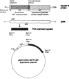
Figure 1
Figure 1. Schematic representation of the strategy for construction of expression vector pGEX- 3X/GC-ANFR-LBD
The synthetic PCR primers (filled arrows on cDNA) were designed to amplify the extracellular ANF-binding domain from the full length murine GC/ANF-R cDNA. The hydrophobic signal sequence (dotted region), the transmembrane domain (vertical hatched region) and the entire cytoplasmic catalytic portion (horizontal hatched region) of the protein were excluded from the region amplified. The BamHI linker restriction sites included in the PCR primers were used to clone the amplified product into the corresponding sites in prokaryotic expression vector pGEX-3X. The resulting expression construct (pGEX-3X/GC- ANFR-LBD) allows the IPTG-inducible expression of the fusion protein with an amino-terminal portion consisting of glutathione-S-transferase gene and the extracellular domain of GC/ANF-R cDNA which is represented by the filled box in the pGEX-3X/GC-ANFR-LBD expression vector.
Figure 2
Figure 2. The expression analysis of the 70 kUa fusion protein consisting of GST and extracellular ANF-binding domain of GC/ANF-R
The bacterial cell lysates were subjected to SDS-PAGE using 7.5% gel. Lane a, cell lysate of transformed E. coli not treated with IPTG; lanes b, c and d, cell lysates of transformed E. coli which were induced with IPTG for 30, 60 and 90 min, respectively, as described in the Materials and Methods. Arrow indicates the position and molecular mass of the 70 kDa fusion protein, induced in a time-dependent manner. Lane e shows the purified 70 kDa fusion protein as a single band after glutathione-affinity chromatography. Bars on the left margin indicate the position and molecular mass of the marker proteins.
Figure 3
Figure 3. Capillary electrophoresis of the purified 70 kDa fusion protein
The samples containing the fusion protein eluted from the glutathione-affinity column were applied on to the capillary column (50 × 75, μID). The electrophoresis was carried out in 50 mM borate buffer, pH 8.5, containing 50 mM SDS at 20 kilo volts. A predominant single protein peak was detected after 8 min at absorbance 280 nm.
Figure 4
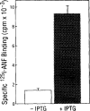
Figure 4. Binding of 125I-ANF to solubilized extracts of bacterial cells transfected with pgex- 3X/GC-ANFR-LBD
The bacterial cells without and with IPTG-treatment were harvested and the cell lysates were prepared as described in the Materials and Methods section. The 50 μl solubilized cell extracts were incubated with 1 nM l25I-ANF in 450 μl binding buffer with and without unlabeled ANF at room temperature for 1 h. The bound l25I-ANF-receptor complex was precipitated by adding 0.25% bovine γ-globulin and 2.5 ml of 10% polyethyleneglycol 8000 as previously described in the Materials and Methods section. The mixture was filtered under vacuum through Whatman GF/B filters, treated with 0.3% (w/v) polyethyleneimine. After washing the free ligand, the radioactivity retained on the filter was counted in Beckman Gamma-5500. The data represent the mean of three separate determinations.
Figure 5
Figure 5. Binding competition of l25I-ANF to the solubilized lysates of bacterial cells transfected with pGEX-3X/GC-ANFR-LBD
The 50 μl lysates from IPTG-induced bacterial cell were incubated with 1 nM l25I-ANF in 450 μl binding buffer for 1 h with various concentrations of unlabeled hormones. The binding assay was carried out as described in the Material and Methods section. •, ANF(99-126); ▲, ANF(102-126); ○, ANF(103-123) and □, angiotensin II and vasopressin.

Similar articles

Cited by

References

    1. de Bold AJ, Borenstein HB, Veress AT, Sonnenberg H. Life Sei. 1981;28:89–94. - PubMed
    1. de Bold AJ. Science. 1985;230:767–770. - PubMed
    1. Cantin M, Genest J. Endocrine Rev. 1985;6:107–127. - PubMed
    1. Inagami T. J Biol Chem. 1989;264:3043–3046. - PubMed
    1. Rosenzweig A, Seidman CE. Ann Rev Biochem. 1991;60:229–255. - PubMed

Publication types

MeSH terms

Substances