. 1993 Jun 3;363(6428):451-4.
doi: 10.1038/363451a0.

Endogenous dynorphins inhibit excitatory neurotransmission and block LTP induction in the hippocampus

Affiliations

Endogenous dynorphins inhibit excitatory neurotransmission and block LTP induction in the hippocampus

J J Wagner et al. Nature. .

Abstract

Although anatomical and neurochemical studies suggest that endogenous opioids act as neurotransmitters, their roles in normal and pathophysiological regulation of synaptic transmission are not defined. Here we examine the actions of prodynorphin-derived opioid peptides in the guinea-pig hippocampus and show that physiological stimulation of the dynorphin-containing dentate granule cells can release endogenous dynorphins, which then activate kappa 1 opioid receptors present in the molecular layer of the dentate gyrus. Activation of kappa 1 receptors by either pharmacologically applied agonist or endogenously released peptide reduces excitatory transmission in the dentate gyrus, as shown by a reduction in the excitatory postsynaptic currents evoked by stimulation of the perforant path, a principal excitatory afferent. In addition, released dynorphin peptides were found to block the induction of long-term potentiation (LTP) at the granule cell-perforant path synapse. The results indicate that endogenous dynorphins function in this hippocampal circuit as retrograde, inhibitory neurotransmitters.


Figures

FIG. 1
FIG. 1
Granule cell responses to molecular layer and hilar stimulation and the effects of U69,593 on excitatory synaptic currents (e.p.s.cs). a, ln a representative cell, e.p.s.cs produced by increasing stimulus intensities of perforant path (20, 25, 30 μA) or hilar (100, 150 μA) stimulation in the presence of bicuculline (10 μM) were both largely blocked by 6-cyano-7-nitroquinoxaline-2,3-dione (CNQX; 5 μM). U69,593 affected neither the membrane conductance of granule cells at membrane potentials from −120 to −60 mV (data not shown), nor the holding current required to clamp the cell at −70 mV. b, In another representative granule cell in the presence of bicuculline, the κ opioid agonist U69,593 (500 nM) inhibited CNQX-sensitive e.p.s.cs from from perforant path. This inhibition was completely reversed by the κ antagonist NBNI (100 nM; n = 4). The slight reduction in e.p.s.c. amplitude in the presence of U69,593 seen after hilar stimulation was neither reversed by NBNI nor statistically significant in 4 replicates (P > 0.05). Individual traces shown in a and b are the averages of two sequential sweeps; the experiment was done 3 times with similar results. Sweep length is 110ms; vertical scales are current amplitudes (pA); calibration is 10ms (horizontal), 40 pA (vertical) for all traces. METHODS. Guinea-pig hippocampal slices (500 μm) were prepared as described and were perfused with Krebs bicarbonate buffer containing (mM): NaCl (125), KCl (3), CaCl2 (2), MgCl2 (1). NaH2P04 (1.25), NaHCO3 (26), and glucose (10), saturated with 95% O2, 5% Co2, pH 7.4. Patch pipettes (3-5 MΩ) contained (mM): CsCl (120), CaCl2 (1), MgCl2 (2), ATP (5), tetraethyl-ammonium-Cl (20), EGTA (10), HEPES (10), adjusted to pH 7.2 with CsOH. Whole-cell voltage clamp recordings were obtained using an Axopatch 200 amplifier and analysed using FastLab software. Uncompensated series resistance was checked throughout the recording period, and the data excluded from analysis if the drift exceeded 20%. To study isolated e.p.s.cs, bicuculline (10 μM) was added to the bath to block GABAA receptors in all studies reported here and CsCl was included in the intracellular recording pipettes to block GABAB receptor-gated potassium currents. Spontaneous activity was virtually eliminated after GABAA receptors were blocked by bicuculline (45 of 45 cells). For statistical analysis we used analysis of variance with or without repeated measures as appropriate. Tukey’s tests were used for post-hoc comparisons; P<0.05 was considered to be significant.
FIG. 2
FIG. 2
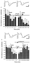
Hilar high-frequency stimulation (HHFS) induces inhibition of perforant path-evoked excitatory postsynaptic potentials (PP e.p.s.cs) recorded in the granule cell, a, HHFS causes a 24% reduction in the amplitude of the granule cell e.p.s.cs. Following recovery (>90% of control) from the initial HHFS, a second HHFS was again able to inhibit PP e.p.s.c. amplitude (compare sweeps 3 and 4). b, HHFS causes a 26% reduction in the PP e.p.s.c. amplitude (compare sweeps 1 and 2). Twelve minutes after addition of 1 μM naloxone to the superfusion buffer, the PP e.p.s.c. amplitude returned to a pre-HHFS level and another HHFS reduced PP e.p.s.c. amplitude by only 4% (compare sweeps 3 and 4). Vertical scales for figure insets are current amplitudes (pA). Stimulus artefacts are truncated. Group data: HHFS significantly decreased e.p.s.c. amplitude at two (−15±2%), three (−13±3%) and four (−16 ±3%) min post-stimulus in normal Krebs bicarbonate buffer (n = 15); it had no effect at any time point when naloxone (1 μM) was added to the perfusate at least 10 min before HHFS (n = 5). METHODS. The opioid-mediated effects of HHFS were monitored by measuring granule cell e.p.s.c. amplitudes evoked by a perforant path test pulse in the presence or absence of naloxone. PP e.p.s.cs were elicited at 0.1 Hz and 6 sweeps averaged into 1 min bins (bars are means ± s.e.m. of the 6 sweeps). For each cell tested, the mean of 3 or 4 min of pre-HHFS e.p.s.c. amplitudes was determined, and the per cent of that control value calculated. Hilar stimulation at high frequency (50 Hz, 1 s train of 0.3 ms, 150-μA pulses) was applied using a concentric bipolar electrode (Kopf, SNE 100). Calibration is 10 ms (horizontal). 25 pA (vertical) for all traces.
FIG. 3
FIG. 3
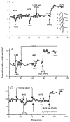
Both norbinaltorphimine and dynorphins antisera block the excitatory effects of hilar high-frequency stimulation on granule cell population spike amplitude and long-term potentiation produced by perforant path high-frequency stimulation on granule cell population spike amplitude and long-term potentiation produced by perforant path high-frequency stimulation (PPHFS). A, Representative experiment showing the effects of HHFS, HHFS followed immediately by PPHFS, and PPHFS on granule cell population spike responses. HHFS (6 1-s, 50 Hz trains of 0.3-ms 300 μA pulses given at a rate of 1 every 10s) decreased spike amplitude by 40% and then 38%. showing nearly identical inhibitory effects of repeated HHFS. Whereas HHFS followed immediately by PPHFS resulted in a minimal change (+2%) in spike amplitude 30 min after stimulation, PPHFS by itself produced a 46% increase 30 min after stimulation (that is, LTP). Inset shows representative traces from a replicate experiment at different times during the protocol: a, control: b,1 min after HHFS; c, 20 min after HHFS; d. 1 min after HHFS immediately followed by PPHFS; and e, 30 min after PPHFS-induced LTP. Responses were evoked at 55 μA (the S1/2, or half-maximal spike amplitude, for this preparation); calibration bars for the inset are 5 ms (horizontal). 1 mV (vertical). B, Effect of 100 nM NBNI on the HHFS-induced attenuation of the population spike and PPHFS-evoked LTP. HHFS produced a 29% decrease in spike amplitude initially, but only an 8% decrease after NBNI was added to the perfusate. Unlike the results shown in A, HHFS given immediately before PPHFS in the presence of NBNI did not block LTP (+31%). C, Effect of dynorphin antisera on HHFS-induced reduction of the population spike and LTP following PPHFS. With normal rabbit serum (1:125) in the perfusate, HHFS decreased spike amplitude 25% (similar in magnitude to A and B). After dynorphin antisera was added to the perfusate, HHFS reduced spike amplitude by only 7% and, unlike the results shown in A, a 22% potentiation was seen with HHFS immediately followed by PPHFS. METHODS. Extracellular recordings were made under the conditions described in Fig. 1 legend, except that the concentrations of CaCl2, and MgCl2 in the extracellular buffer were each increased to 4 mM to inhibit hyperexcitability in the presence of the 10 μM of bicuculline. Recording pipettes were filled with 3 M NaCl. The perforant path stimulation-induced dentate granule cell population response amplitude was measured from peak to peak using a digitizing oscilloscope. A stimulus intensity was chosen that evoked a half-maximal granule cell population spike amplitude (S1/2). The LTP induction paradigm consisted of a total of 9 pulses (0.3 ms. 300 μA) to the perforant path given as three trains (each with three stimuli) applied at 10-s intervals with 10 ms between stimuli. LTP was assessed at 25–30 min post-PPHFS. Pooled dynorphin antisera included rabbit polyclonal antisera raised against dynorphin A1–8, dynorphin B and α-neoendorphin. The preparation and characterization of this antisera has been described6.30. All three antisera were added to the perfusate at the following dilutions: 1:300, dynorphin A1–8; 1:250. dynorphin B; 1:1,000, α-neoendorphin (for a final litre of 1:120 serum in the medium).

References

    1. Gall C, Brecha N, Karten HJ, Chang K. J comp Neurol. 1981;198:335–350. - PubMed
    1. McGinty JF, Henriksen SJ, Goldstein A, Terenius L, Bloom FE. Proc natn Acad Sci USA. 1983;80:589–593. - PMC - PubMed
    1. Chavkin C, Shoemaker WJ, McGinty JF, Bayon A, Bloom FE. J Neurosci. 1985;5:808–816. - PMC - PubMed
    1. McLean S, Rothman R, Jacobson A, Rice K, Herkenham M. J comp Neurol. 1987;255:497–510. - PubMed
    1. Chavkin C, Bakhit C, Weber E, Bloom FE. Proc natn Acard Sci USA. 1983;80:7669–7673. - PMC - PubMed

Publication types

MeSH terms