. 1993 Oct 14;365(6447):666-9.
doi: 10.1038/365666a0.

A nuclear localization signal within HIV-1 matrix protein that governs infection of non-dividing cells

Affiliations

A nuclear localization signal within HIV-1 matrix protein that governs infection of non-dividing cells

M I Bukrinsky et al. Nature. .

Abstract

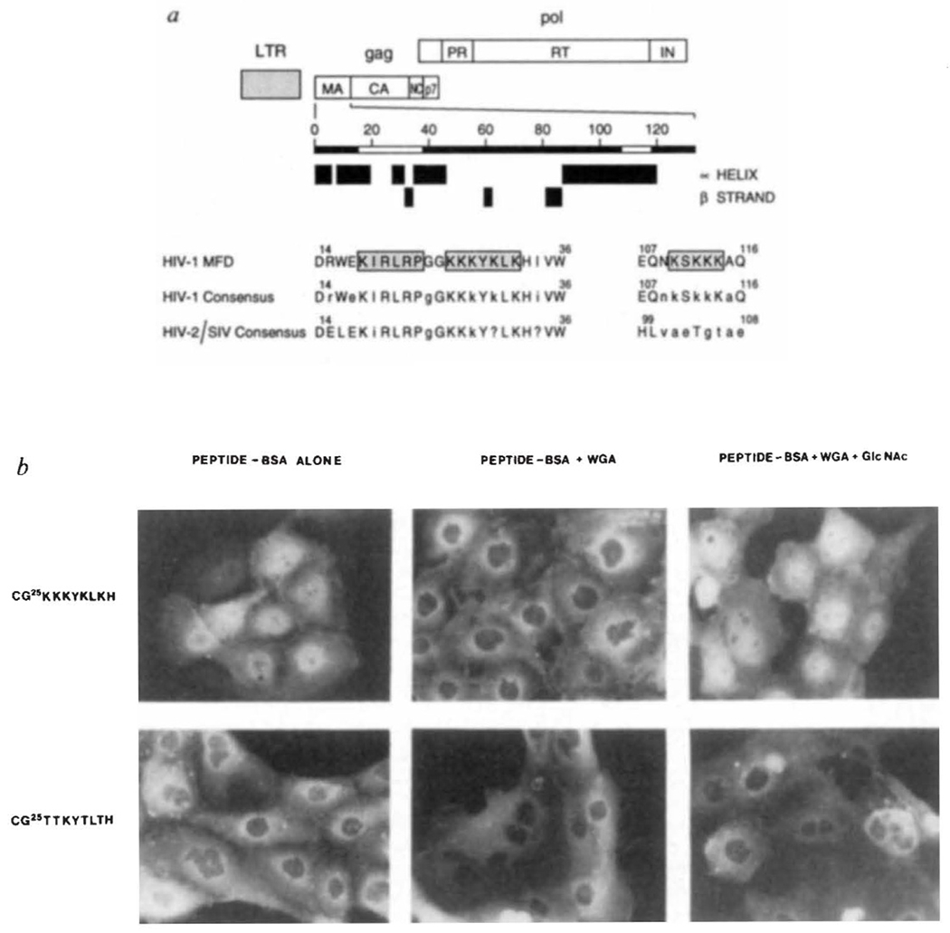
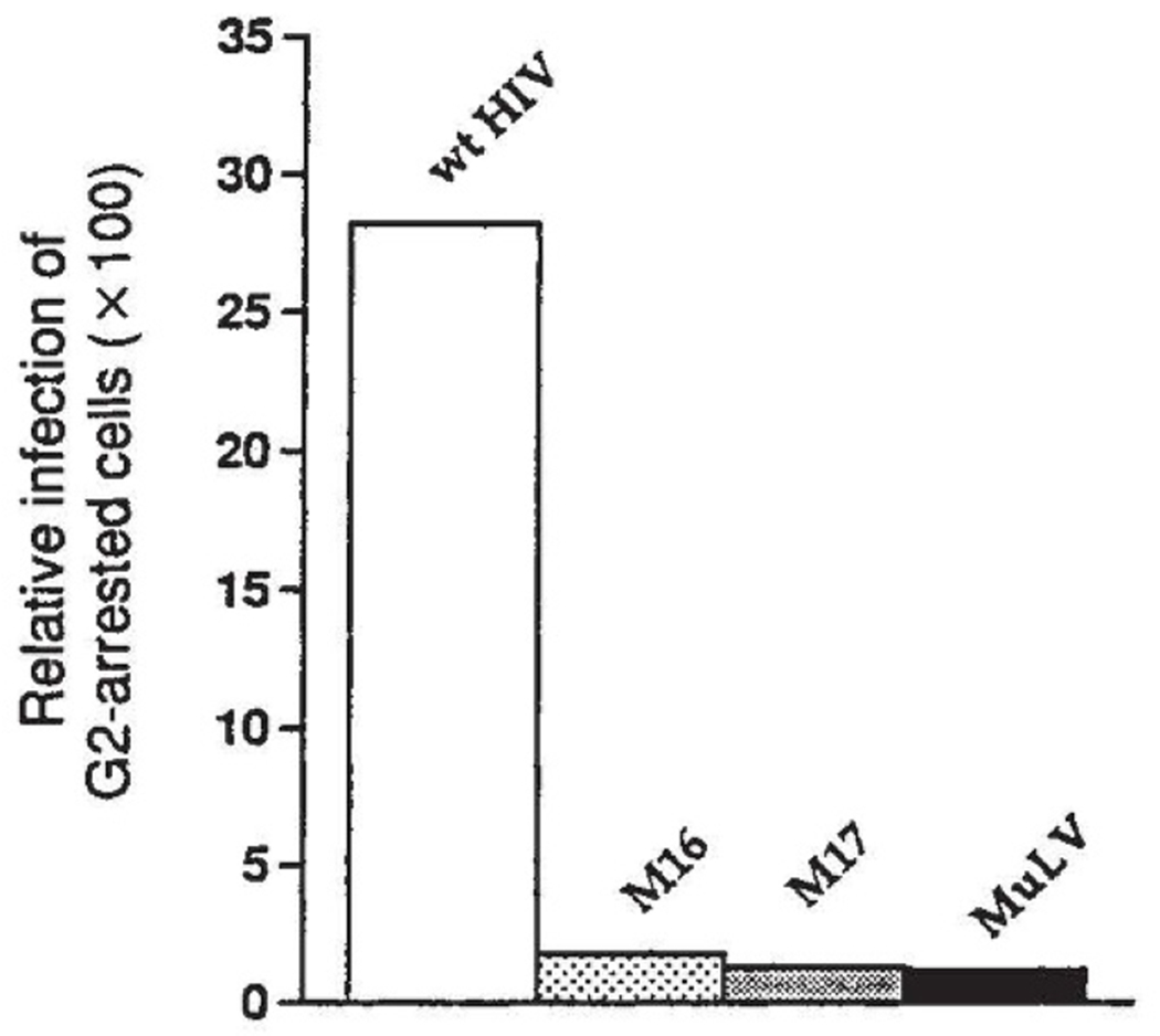
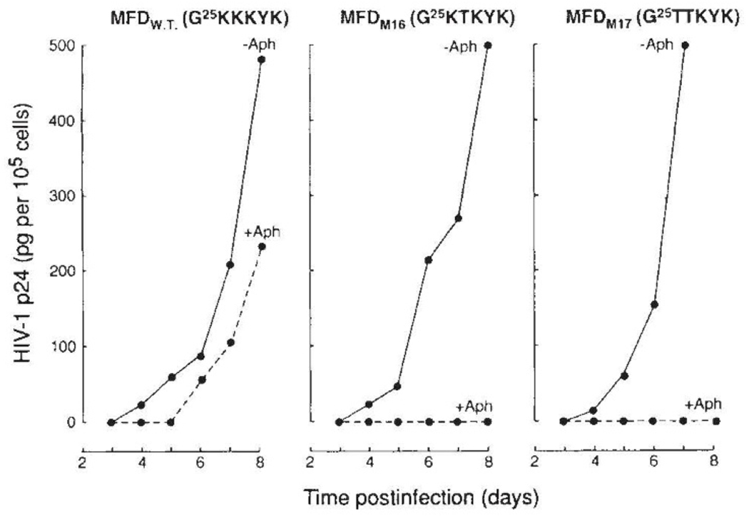
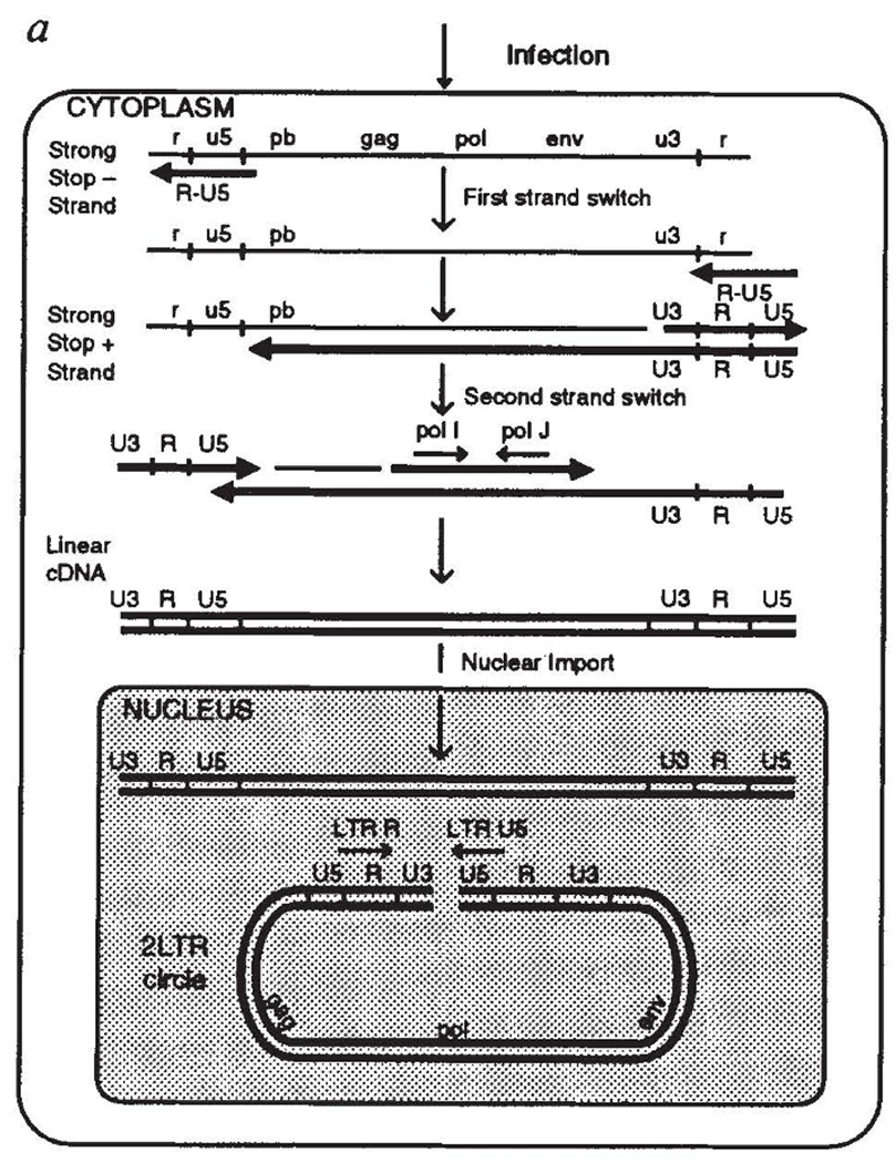
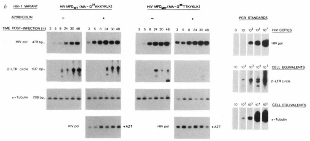
Permissiveness of the host cell to productive infection by oncoretroviruses is cell-cycle dependent, and nuclear localization of viral nucleoprotein preintegration complexes will occur only after cells have passed through mitosis. In contrast, establishment of an integrated provirus after infection by the lentivirus HIV-1 is independent of host cell proliferation. The ability of HIV-1 to replicate in non-dividing cells is partly accounted for by the karyophilic properties of the viral preintegration complex which, after virus infection, is actively transported to the host cell nucleus. Here we report that the gag matrix protein of HIV-1 contains a nuclear localization sequence which, when conjugated to a heterologous protein, directs its nuclear import. In addition, HIV-1 mutants containing amino-acid substitutions in this nuclear localization signal integrate and replicate within dividing but not growth-arrested cells, and thus display a phenotype more representative of an oncoretrovirus.


Figures

FIG. 1
FIG. 1
Putative nuclear targeting sequences within the HIV-1 gag matrix protein. a, Boundaries of the major products of the gag and pol polyproteins are indicated. The positions of predicted α-helices and β-strands were determined as in ref. . Sequences that resemble common NLS motifs are boxed and stippled. Amino-acid numbering, alignment and description of consensus sequences was helped by information contained within the HIV-1 sequence data base. Upper case letters represent invariant residues whereas conserved and non-conserved amino acids are denoted by lower case letters and the question mark, respectively. b, Subcellular location of HIV-1 matrix NLS peptide–BSA conjugates after microinjection into rat epithelial cells. METHODS. PtK1 cells (rat kangaroo kidney epithelia) were grown on glass cover slips in F-12 medium supplemented with 10% fetal bovine serum. Cells were microinjected in Hank’s balanced salt solution, essentially as detailed elsewhere. Fluoresceinated HIV-1 matrix peptide–BSA conjugates containing an average of 6 peptides per BSA molecule were injected with or without WGA alone (2.5 mg ml−1) or WGA and its ligand, GlcNAc(0.5 M). Microinjected cells were incubated for 30 min at 37 °C, fixed and photographed.
FIG. 2
FIG. 2
Relative infection of G2-arrested HeLa CD4 cells by HIV-1, MuLV and HIV-1 NLS matrix mutants. HeLaCD4-LTR/βgal cells were arrested in G2 by γ-irradiation, and infection was assayed by transactivation of an endogenous LTR–β-galactosidase gene as described. In this assay, HeLa cells, which have been engineered to express high levels of the HIV receptor, CD4, contain a single copy of the bacterial β-galactosidase gene under transcriptional control of the HIV-1 LTR. On HIV-1 infection and integration β-galactosidase is produced after transactivation of the LTR by tat protein. Infected cells are scored after visualization of β-galactosidase activity in situ by staining cells with x-gal (5-bromo-4-chloro-3-indolyl-β-d-galactopyranoside). The relative infection of G2-arrested cells is the number of infected cells in the γ-irradiated (non-proliferating) culture divided by the number of infected cells in the proliferating culture for each viral infection. wt HIV is the parental MFD strain; M16 is the single Lys 27→Thr matrix mutant of HIV-1 MFD; M17 is the double Lys 26, 27→Thr mutant of HIV-1 MFD; and MuLV is a MuLV-based retrovirus vector that encodes the HIV-1 tat gene. METHODS. Amino-acid substitutions within HIV-1 gag matrix were introduced by site-directed mutagenesis. A 3-kilobase (kb) Sphl fragment of the infectious molecular clone, HIV-1 MFD (similar in genotype to the prototypic HXB2 isolate), was inserted in M13mp19. Single (Lys 27→Thr) and double (Lys 26, 27→Thr) amino-acid substitutions were created by annealing M13 phage DNA with the mutagenic oligonucleotides, M16(5′-aggcctgggggaaagacaaaatat-3′) and M17(5′-aggcctgggggaacgacaaaatat-3′), respectively. Both oligonucleotides were designed to introduce translationally silent Stul recognition sites. DNA fragments containing the desired mutations were inserted in the HIV-1 MFD DNA backbone. Wild-type and mutant HIV-1 variants were recovered 48 h after transfection of HeLa cells and quantified by HIV-1 gag p24 ELISA. When cells are transfected with infectious DNA clones, events preceding provirus establishment are bypassed and only late events including assembly and release of virion particles are reconstituted. The amino-acid substitutions within the HIV-1 gag matrix NLS did not influence virus maturation and both quantity and morphology of virus particles produced from HeLa cells transfected with matrix mutant and wild-type molecular clones of HIV-1 were identical (not shown).
FIG. 3
FIG. 3
Replication of wild-type and gag matrix mutants of HIV-1 in dividing and in growth-arrested CD4+ cells. CD4+ MT-4 cell cultures were arrested at the G1/S interface of the cell cycle by the action of aphidicolin as detailed elsewhere. Aphidicolin was removed 72 h after infection with HIV-1 (50 pg per 106 cells). At 24-h intervals, 1 × 10⁵ cells were removed for quantification of cell-associated HIV-1 gag p24 in proliferating (solid line) and in growth-arrested (hatched line) cultures.
FIG. 4
FIG. 4
Synthesis and nuclear import of viral DNA by wild-type and gag matrix mutants of HIV-1 in proliferating and growth-arrested CD4+ T-cell cultures. a, Major reverse transcription products in cytoplasm and nucleus of HIV-1-infected cells. After reverse transcription is initiated, two strand-switching events result in a full-length cDNA containing duplicated U3, R and U5 regions. After transport to the nucleus, some linear molecules undergo circularization, yielding 2 LTR circle forms. Although such circle forms of viral DNA do not appear to be precursors of the provirus, they are formed only after synthesis of full-length viral cDNA and its transport to the nucleus, and thus represent a specific marker with which to follow nuclear localization of viral DNA. HIV-1 pol I and J primers amplify products of reverse transcription which follow the second strand switch, whereas LTR R and U5 primers amplify 2 LTR circle forms of extrachromosomal viral DNA. Viral RNA and cDNA are represented by thin lines/lower-case letters and thick lines/uppercase letters, respectively. pb, Primer binding site. b, Proliferating (− Aph) and growth-arrested ( +Aph) CD4+ MT-4 cells were infected with HIV-1 as outlined in Fig. 3. At the indicated times postinfection, cell aliquots were withdrawn (2–3 × 10⁵ cells) for isolation of total cellular DNA as detailed elsewhere. HIV-1 pol-specific amplification products obtained in the presence of AZT (1 μm) represent incomplete reverse transcription products contained within the virion and not de novo synthesis of viral DNA,. METHODS. PCR conditions for amplification of linear and circle forms of viral DNA were exactly as detailed elsewhere,. One and two rounds of PCR were used for pol and LTR circle primers, respectively. PCR amplification products were visualized after Southern blot transfer and hybridization with [α-32P]-labelled oligonucleotide probes. HIV-1 pol standards were generated by PCR on log dilutions of cloned HIV-1 MF DNA whereas standards for circle viral DNA forms and cellular DNA generated by PCR on log dilutions of HIV-1-infected MT-4 cells.
FIG. 4
FIG. 4
Synthesis and nuclear import of viral DNA by wild-type and gag matrix mutants of HIV-1 in proliferating and growth-arrested CD4+ T-cell cultures. a, Major reverse transcription products in cytoplasm and nucleus of HIV-1-infected cells. After reverse transcription is initiated, two strand-switching events result in a full-length cDNA containing duplicated U3, R and U5 regions. After transport to the nucleus, some linear molecules undergo circularization, yielding 2 LTR circle forms. Although such circle forms of viral DNA do not appear to be precursors of the provirus, they are formed only after synthesis of full-length viral cDNA and its transport to the nucleus, and thus represent a specific marker with which to follow nuclear localization of viral DNA. HIV-1 pol I and J primers amplify products of reverse transcription which follow the second strand switch, whereas LTR R and U5 primers amplify 2 LTR circle forms of extrachromosomal viral DNA. Viral RNA and cDNA are represented by thin lines/lower-case letters and thick lines/uppercase letters, respectively. pb, Primer binding site. b, Proliferating (− Aph) and growth-arrested ( +Aph) CD4+ MT-4 cells were infected with HIV-1 as outlined in Fig. 3. At the indicated times postinfection, cell aliquots were withdrawn (2–3 × 10⁵ cells) for isolation of total cellular DNA as detailed elsewhere. HIV-1 pol-specific amplification products obtained in the presence of AZT (1 μm) represent incomplete reverse transcription products contained within the virion and not de novo synthesis of viral DNA,. METHODS. PCR conditions for amplification of linear and circle forms of viral DNA were exactly as detailed elsewhere,. One and two rounds of PCR were used for pol and LTR circle primers, respectively. PCR amplification products were visualized after Southern blot transfer and hybridization with [α-32P]-labelled oligonucleotide probes. HIV-1 pol standards were generated by PCR on log dilutions of cloned HIV-1 MF DNA whereas standards for circle viral DNA forms and cellular DNA generated by PCR on log dilutions of HIV-1-infected MT-4 cells.

Comment in

References

    1. Varmus HE & Swanstrom R in RNA Tumor Viruses (eds Weiss R, Teich N, Varmus HE & Coffin J) 369–512 (Cold Spring Harbor Laboratory Press, New York, 1984).
    1. Roe T, Reynolds TC, Yu G & Brown PO EMBO J. 12, 2099–2108 (1993). - PMC - PubMed
    1. Weinberg JB, Mathews TJ, Cullen BR & Malim MH J. exp. Med 174, 1477–1482 (1991). - PMC - PubMed
    1. Bukrinsky MI et al. Proc. natn. Acad. Sci. U.S.A 89, 6580–6584 (1992). - PMC - PubMed
    1. Lewis P Hensel M &, Emerman M EMBO. J 11, 3053–3068 (1992). - PMC - PubMed

Publication types

MeSH terms