

Structure of the VH and VL segments of monoreactive and polyreactive IgA autoantibodies to DNA in patients with systemic lupus erythematosus
- PMID: 8144908
- PMCID: PMC4631053
Structure of the VH and VL segments of monoreactive and polyreactive IgA autoantibodies to DNA in patients with systemic lupus erythematosus
Abstract
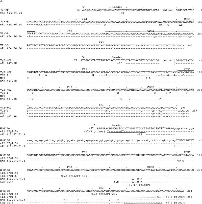
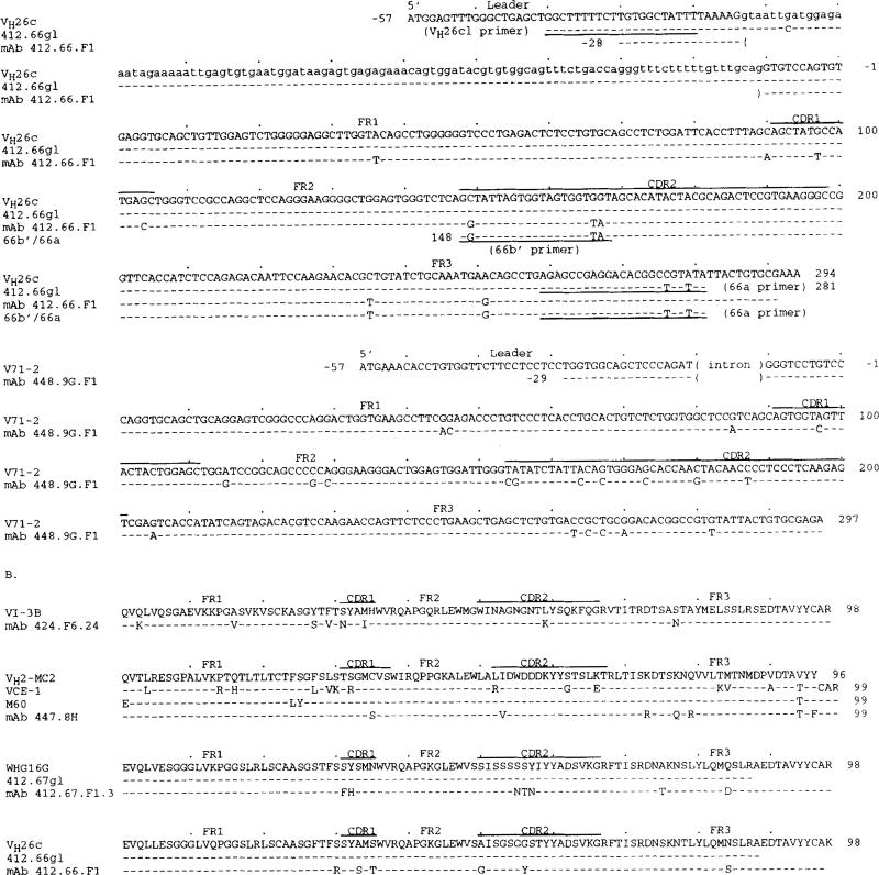
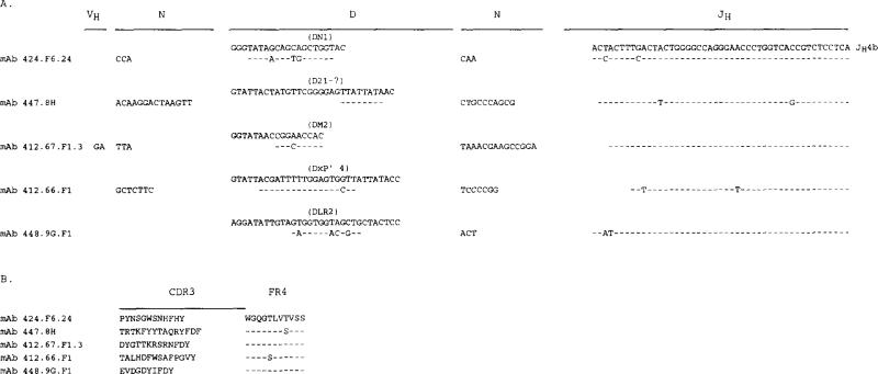
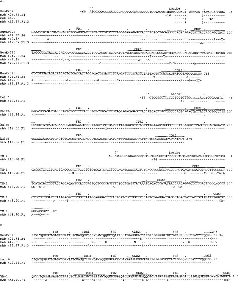
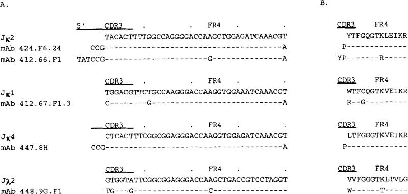
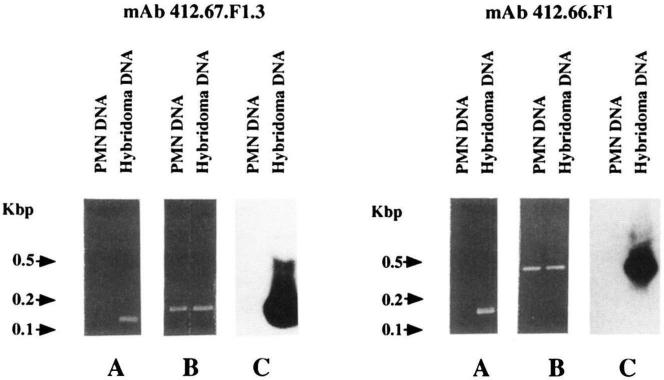
Anti-DNA IgA autoantibodies play an important immunopathologic role in SLE patients. To analyze the cellular origin and the VH and VL structure of anti-DNA IgA autoantibodies, we generated five IgA1 mAbs to DNA using B lymphocytes from three SLE patients. Two mAbs bound to ssDNA only and one to both ssDNA and dsDNA (monoreactive antibodies). The remaining two mAbs bound to DNA (one to ssDNA and the other to both ssDNA and dsDNA) and to other self and foreign Ag (polyreactive antibodies). The IgA mAb relative avidity for DNA ranged from 7.5 x 10(-8) to 8.0 x 10(-10) g/microliters. The anti-DNA IgA mAb used VH segments of the VHI(VI-3b), VHII (VH2-MC2), VHIII (WHG16G and VH26c), and VHIV (V71-2) families in conjunction with V kappa I, V kappa IIIb, or V lambda I segments. All IgA mAb VH segments were juxtaposed with JH4b segments. The heavy chain CDR3 sequences were divergent in composition and length. When compared with those of the closest reported germ line genes, the IgA mAb VH and VL gene sequences displayed a number of differences. That these differences represented somatic point mutations was formally proved in both the monoreactive IgA mAb 412.67.F1.3 and the polyreactive IgA mAb 412.66.F1 VH segments by differential PCR amplification and cloning and sequencing of genomic DNA from the mAb-producing cell lines and autologous polymorphonuclear cells. The sequences of the germ line genes that putatively gave rise to the mAb 412.67.F1.3 and mAb 412.66.F1 VH segments were identical with those of the WHG16G and VH26c genes, respectively. In not only the monoreactive mAb 412.67.F1.3 but also the polyreactive mAb 412.66.F1 and mAb 448.9G.F1 VH segments, the higher concentration of replacement (R) mutations and the higher R:S (silent) mutation ratios in the complementarity-determining region (infinity; 19:0) than in the framework region (1.0) (p = 0.00001, chi 2 test) were highly consistent with selection by Ag. In the five IgA mAb VH and VL segments, the putative and verified somatic point mutations yielded 68 amino acid replacements, of which 38 were nonconserved. Twenty of these yielded positively charged or polar residues that play a major role in DNA binding, including seven Arg, five Lys, three Tyr, two Gln, two His, and a Thr. The conserved amino acid changes included seven Asn. These findings suggest that anti-DNA IgA autoantibodies use a broad selection of VH and VL genes and enhance their fit for Ag by undergoing somatic hypermutation and Ag selection.(ABSTRACT TRUNCATED AT 400 WORDS)
Figures
Similar articles
-
Structural analysis of the VH-D-JH segments of human polyreactive IgG mAb. Evidence for somatic selection.J Immunol. 1993 Oct 1;151(7):3604-16. J Immunol. 1993. PMID: 8376796 Free PMC article.
-
VH and V kappa segment structure of anti-insulin IgG autoantibodies in patients with insulin-dependent diabetes mellitus. Evidence for somatic selection.J Immunol. 1994 Feb 1;152(3):1430-41. J Immunol. 1994. PMID: 8301143 Free PMC article.
-
Clonal analysis of a human antibody response. II. Sequences of the VH genes of human IgM, IgG, and IgA to rabies virus reveal preferential utilization of VHIII segments and somatic hypermutation.J Immunol. 1993 Feb 15;150(4):1325-37. J Immunol. 1993. PMID: 8432980 Free PMC article.
-
B-1 cellular origin and VH segment structure of IgG, IgA, and IgM anti-DNA autoantibodies in patients with systemic lupus erythematosus.Ann N Y Acad Sci. 1995 Sep 29;764:410-23. doi: 10.1111/j.1749-6632.1995.tb55856.x. Ann N Y Acad Sci. 1995. PMID: 7486556 Review. No abstract available.
-
The role of germline gene expression and somatic mutation in the generation of autoantibodies to DNA.Mol Immunol. 1990 Mar;27(3):203-10. doi: 10.1016/0161-5890(90)90131-i. Mol Immunol. 1990. PMID: 2188119 Review.
Cited by
-
A humanized mouse that mounts mature class-switched, hypermutated and neutralizing antibody responses.Nat Immunol. 2024 Aug;25(8):1489-1506. doi: 10.1038/s41590-024-01880-3. Epub 2024 Jun 25. Nat Immunol. 2024. PMID: 38918608 Free PMC article.
-
Is There a Role for Natural Antibodies in Rejection Following Transplantation?Transplantation. 2019 Aug;103(8):1612-1619. doi: 10.1097/TP.0000000000002743. Transplantation. 2019. PMID: 30951015 Free PMC article. Review.
-
Analysis of the structural correlates for antibody polyreactivity by multiple reassortments of chimeric human immunoglobulin heavy and light chain V segments.J Exp Med. 1994 Sep 1;180(3):885-95. doi: 10.1084/jem.180.3.885. J Exp Med. 1994. PMID: 8064239 Free PMC article.
-
Nature and functions of autoantibodies.Nat Clin Pract Rheumatol. 2008 Sep;4(9):491-8. doi: 10.1038/ncprheum0895. Nat Clin Pract Rheumatol. 2008. PMID: 18756274 Free PMC article. Review.
-
Determination of gene organization in individual haplotypes by analyzing single DNA fragments from single spermatozoa.Proc Natl Acad Sci U S A. 1998 Sep 1;95(18):10791-6. doi: 10.1073/pnas.95.18.10791. Proc Natl Acad Sci U S A. 1998. PMID: 9724783 Free PMC article.
References
-
- Tan EM. Antinuclear antibodies: diagnostic markers for autoimmune diseases and probes for cell biology. Adv. Immunol. 1989;44:93. - PubMed
-
- Zouali M, Stollar BD, Schwartz RS. Origin and diversification of anti-DNA antibodies. Itnmunol. Rev. 1988;105:137. - PubMed
-
- Nakamura M, Burastero SE, Ueki Y, Larrick JW, Notkins AL, Casali P. Probing the normal and autoimmune B cell repertoire with Epstein-Barr virus: frequency of B cells producing monoreactive high affinity autoantibodies in patients with Hashimoto's disease and systemic lupus erythematosus. J. Immunol. 1988;141:4165. - PubMed
-
- Casali P, Burastero SE, Balow JE, Notkins AL. High affinity antibodies to ssDNA are produced by CD5− B cells in SLE patients. J. Immunol. 1989;143:3476. - PubMed
Publication types
MeSH terms
Substances
Associated data
- Actions
- Actions
- Actions
- Actions
- Actions
- Actions
- Actions
- Actions
- Actions
- Actions
Grants and funding
LinkOut - more resources
Full Text Sources
Other Literature Sources
Medical
Miscellaneous