. 1993 Jun;118(2):377-88.
doi: 10.1242/dev.118.2.377.

Function and spatial distribution in developing chick retina of the laminin receptor alpha 6 beta 1 and its isoforms

Affiliations

Function and spatial distribution in developing chick retina of the laminin receptor alpha 6 beta 1 and its isoforms

I de Curtis et al. Development. 1993 Jun.

Abstract

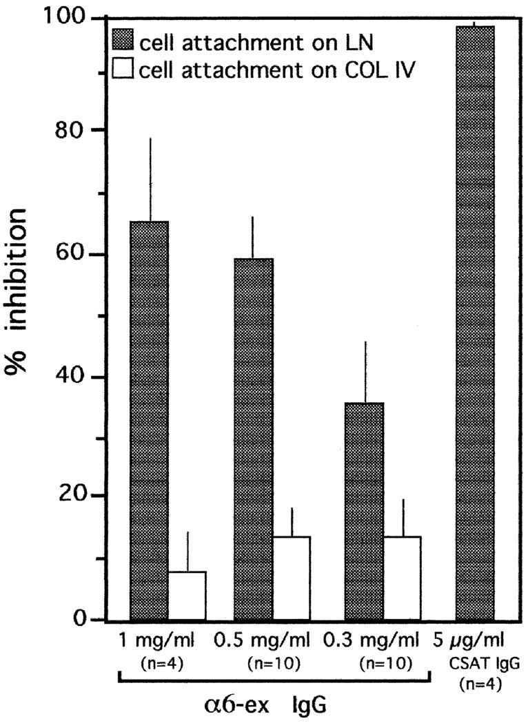
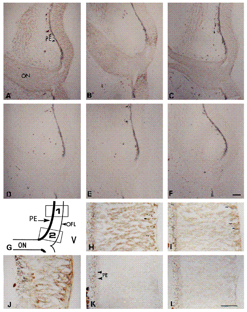
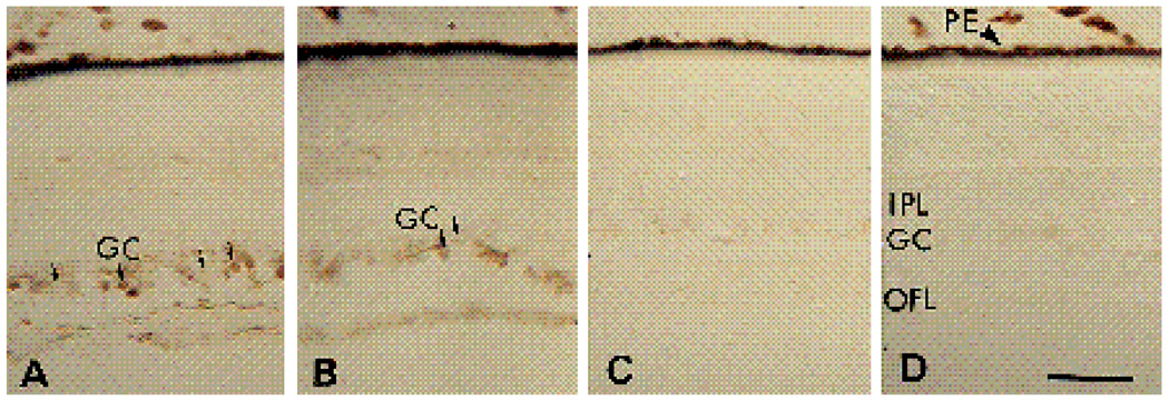
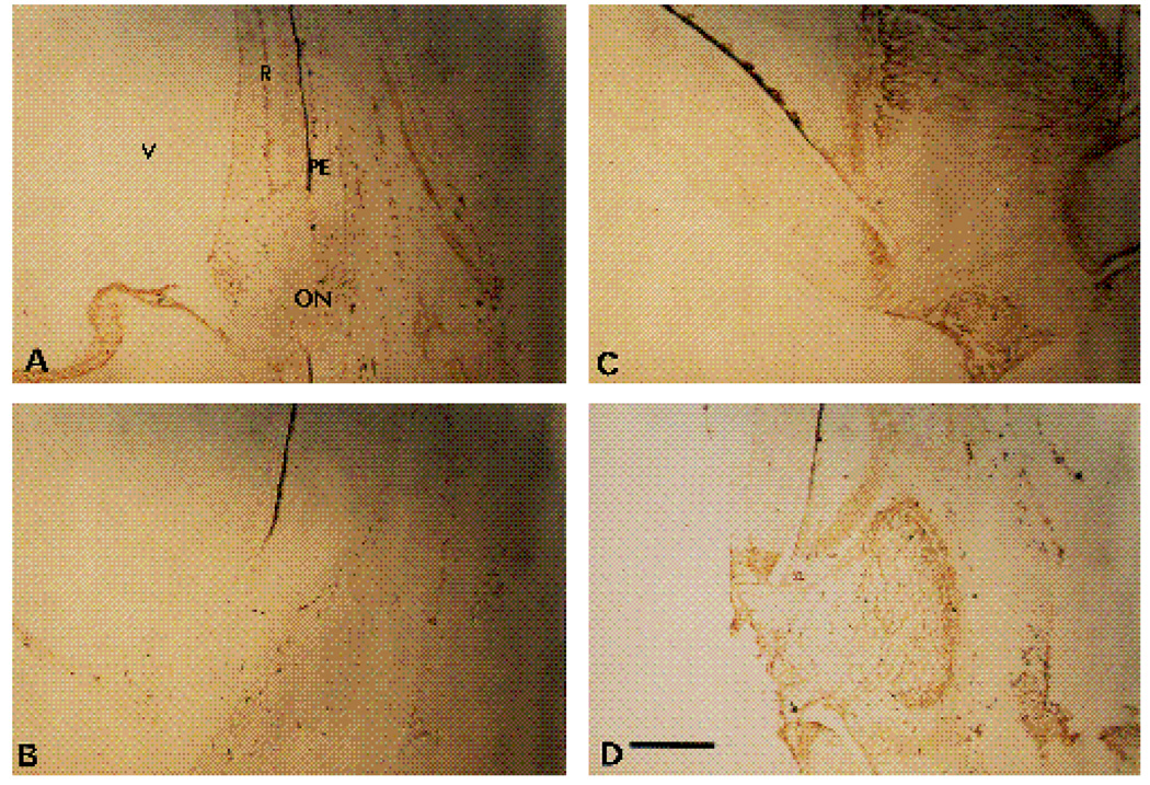
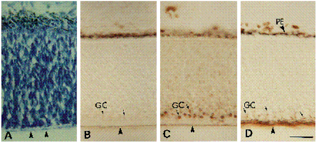
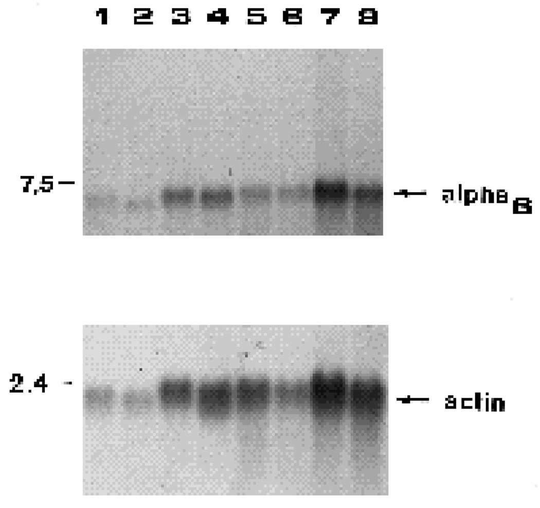
We have recently shown that the laminin-binding integrin receptor, alpha 6 beta 1, is prominently expressed in the developing chick retina, and its expression and activity are regulated during development on both retinal ganglion cells and other neural retinal cells. In the present study, we show that antibodies specific for the extracellular portion of the chick alpha 6 subunit dramatically inhibit interactions in vitro between embryonic day 6 neural retinal cells and laminin, showing that alpha 6 beta 1 functions as an important laminin receptor on developing retinal neurons. In previous work, we showed that alpha 6 mRNA levels on retinal ganglion cells decrease dramatically after E6 during the period that RGC axons innervate the optic tectum. In the present study, we show decreases in alpha 6 mRNA are not prevented by ablation of the optic tectum, indicating that tectal contact is not the major cause of this decrease. Within the embryonic retina, the alpha 6 subunit is codistributed, in part, with laminin, suggesting that it functions as a laminin receptor during retina development in vivo. Furthermore, two isoforms of the alpha 6 protein with distinct cytoplasmic domains generated by differential splicing have quite different distribution patterns in the retina, suggesting that these two isoforms may have different functions during retinal development.


Figures

Fig. 1
Fig. 1
Sequence comparisons between human and chick cytoplasmic domains of integrin α6 proteins. In A, the entire amino acid sequences of the chick (top) and human (bottom) A class cytoplasmic domains are aligned. Note that 34 of 35 amino acid residues are identical. In B, the most 3′ sequenced portion of the chick α6 mRNA is aligned with the same region in the human α6 mRNA. Note the strong homology at the nucleic acid level. Note also that an open reading frame in the chick sequence (top) is very similar to an open reading frame in the human sequence (bottom), which has been shown to constitute the cytoplasmic domain of the B-isoform of the human α6 subunit (Tamura et al., 1991). Only the amino terminal portion of this reading frame is present in sequenced chick cDNAs and is aligned in this figure. Vertical lines indicate that identical amino acids that are present in each sequence.
Fig. 2
Fig. 2
Immunoprecipitations from a cell lysate of metabolically labeled E6 retinal neurons. Cell lysates were prepared as described in the Experimental Procedures. For each lane, the same amount of TCA-precipitable radioactivity was used for immunoprecipitation with α6-EX (lane 1), α6-cytoA (lane 2), α6-cytoB (lane 3), or chick β1 (lane 4) polyclonal antibodies.
Fig. 3
Fig. 3
Cell attachment assay of E6 retinal neurons on laminin (LN) or collagen IV (COL IV). Cells were prepared by trypsinization of E6 retinae and the cell attachment assay was performed for 1 hour at 37°C as described in the Experimental Procedures. Results of experiments with a function blocking β1 subunit-specific mAb (CSAT) and α6EX IgG are shown. In every experiment, each sample was tested in triplicate. Bars indicate the standard error; n represents the number of experiments.
Fig. 4
Fig. 4
Neurite outgrowth of E6 retinal neurons on LN (A–C) or COL IV (D–F). Retinal neurons were prepared from E6 retinae and used in the neurite outgrowth assay as described in the Materials and methods. The cells were cultured overnight without IgG (A,D), with 0.5 mg/ml α6-EX IgG (B,E), or with 0.5 mg/ml normal rabbit IgG (C,F). Bar, 40 µm.
Fig. 5
Fig. 5
Micrographs of immunoperoxidase staining on E6 chick retina. 16 µm sections of E6 retina were incubated with α6-EX (A,H), α6-cytoA (B,J,K), or α6-cytoB (C,I) antibodies. D,E and Fare control stainings with preimmune IgG, for A,B and C, respectively. L is the control staining with preimmune IgG for J and K. G shows a diagram of the low power magnification of the retina as shown in panels A–F. Box 1 indicates the area of the retina shown in panels H,I,K and L. Box 2 indicates the area of the retina shown in J. ON, optic nerve; PE, pigmented epithelium; OFL, optic fiber layer; V, vitreous. Bars are 20 µm in A–F, 10 µm in H–L.
Fig. 6
Fig. 6
Micrographs of immunoperoxidase staining on E12 chick retina. 16 µm sections were stained with α6-ex (A), α6-cytoB (B) and α6-cytoA (C) antibodies. D is a section stained with preimmune IgG for the α6-cytoA antibody. IPL, inner plexiform layer; OFL, optic fiber layer; PE, pigmented epithelium. Arrows point to examples of cells with strong or faint immunoperoxidase reaction product in the ganglion cell layer. Bar, 10 µm.
Fig. 7
Fig. 7
Low power micrographs of α6 and α6A expression patterns in E12 chick retina. 16 µm sections were stained with α6EX IgG (A), preimmune IgG for α6EX (B), or α6cytoA (C and D). The region surrounding the optic nerve is visible in each panel. ON, optic nerve; PE, pigment epithelium; V, vitreous. Reaction product is brown. Note localized distribution of α6A in retina adjacent to the optic nerve, within the optic nerve, and in tissues surrounding the retina. Bar, 10 µm.
Fig. 8
Fig. 8
Micrographs of E6 (A–D) 16 µm thick retina sections stained with thionin (A) or with immunoperoxidase after incubation with anti-LN (B), α6-EX (C), or G4 (D) IgG. GC, ganglion cells (arrows); PE, pigmented epithelium. Arrowheads indicate the border between the optic fiber layer and the vitreous. Bar, 10 µm
Fig. 9
Fig. 9
Effects of tectal ablation on integrin α6 mRNA levels in retinal ganglion cells and other retinal cells. Retinal ganglion cells (RGC) and retinal ganglion cell depleted fractions (non-RGC) were used to prepare total RNA samples. Lanes 1–4 are RNA samples from one experiment; lanes 5–8 are RNA samples from a second experiment. RGC, controls (lanes 1,5); RGC, tectum-ablated (lanes 2,6); non-RGC, controls (lanes 3,7); non-RGC, tectum-ablated (lanes 4,8). RNA samples were fractionated by agarose gel electrophoresis, transferred to nitrocellulose and incubated with 32P-α6 probe. The upper section of this blot was incubated with the 32P-α6 probe, the lower section with the 32Pactin probe. The positions of DNA standards are indicated in kb on the left. When quantitated, tectal ablation resulted in 15% and 47% increases in α6 mRNA in the RGC fractions, and 11% and 16% decreases in the non-RGC fractions respectively in the two experiments depicted here.

References

    1. Adler R, Jerdan J, Hewitt AT. Responses of cultured neural retinal cells to substratum-bound laminin and other extracellular matrix molecules. Dev. Biol. 1985;112:100–114. - PubMed
    1. Bodary SC, Napier MA, McLean JW. Expression of recombinant platelet glycoprotein IIb/IIIa results in a functional fibrinogen-binding complex. J. Biol. Chem. 1989;264:18859–18862. - PubMed
    1. Bottenstein JE, Skaper SD, Varon S, Sato J. Selective survival of neurons from chick embryo sensory ganglionic dissociates by use of a defined, serum-free medium. Exp. Cell Res. 1980;125:183–190. - PubMed
    1. Carter WG, Kaur P, Gil SG, Gahr PJ, Wayner EA. Distinct functions for integrins α3β1 in focal adhesions and α6β4/bullous pemphigoid antigen in a new stable anchoring contact (SAC) of keratinocytes: Relation to hemidesmosomes. J. Cell Biol. 1990a;111:3141–3154. - PMC - PubMed
    1. Carter WG, Wayner EA, Bouchard TS, Kaur P. The role of integrins α2β1 and α3β1 in cell-cell and cell-substrate adhesion of human epidermal cells. J. Cell Biol. 1990b;110:1387–1404. - PMC - PubMed

Publication types