. 1993;3(2):163-74.

Molecular organization of the rat glia-derived nexin/protease nexin-1 promoter

Affiliations

Molecular organization of the rat glia-derived nexin/protease nexin-1 promoter

H Ernø et al. Gene Expr. 1993.

Abstract

The first three exons and the promoter of rat glia-derived nexin, also called protease nexin-1 (GDN/PN-1), have been identified through analysis of rat genomic clones. A 1.6 kilobase (kb) fragment containing 105 base pairs of the first exon and 5'-flanking sequences was sequenced. The 5'-flanking sequence and the first exon were found to be GC-rich, indicating that the 5' region of the rat GDN/PN-1 gene resides within a CpG island. A TATA box-like sequence, but no CAAT box, was found. The rat GDN/PN-1 promoter contains five SP1 consensus sites, four consensus sites for the MyoD1 transcription factor, and one binding site for the transcription factors NGFI-A, NGFI-C, Krox-20, and Wilms tumor factor. The presence of these consensus sequences is consistent with the known expression pattern of GDN/PN-1. Primer extension and RNase protection assays identified one transcriptional start site. The 1.6 kb promoter fragment cloned in a reporter plasmid was found to induce firefly luciferase expression in a cell-specific manner. A positive regulatory element is localized in the region -1545 to -389. In vitro CpG methylation blocked transcription from the GDN/PN-1 promoter in rat hepatoma cells but not in C6 rat glioma cells.


Figures

Figure 1
Figure 1
Maps of probes and isolated genomic clones. Open boxes represent 5′ noncoding sequences, filled boxes coding sequences. Exons are not drawn to scale. A. The 280 bp probe used for isolating the genomic clones containing the second and third exons of rat GDN/PN-1, and localization of the 83 nt and 9 nt oligonucleotides used for preparing the probe to isolate the rat GDN/PN-1 promoter clones. The asterisk shows the position of the splice site. B. Restriction map of isolated genomic clones, b: BamH I sites; e: EcoR I sites; h: Hind III; and s: Sal I sites. The localization of the primers used for primer extension are indicated by arrows. a is primer “exon II.1,” b is primer “intron I.1,” c is primer “exon I.1,” and d is primer “exon I.2.”
Figure 2
Figure 2
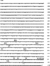
Sequence of rat GDN/PN-1 promoter. The sequence shown extends from the internal BamH I site in the first exon to the upstream EcoR I site shown in Figure 1B. The transcriptional start site is indicated by an arrow; the TATA box, Sp1, AP-2, and MyOD sites are shown in boldface. Sp1/NGFI/Sp1 indicates the overlapping bindings sites for Sp1 and NGFI-A/C, Krox-20, and Wilms tumor factor; the latter binding site is in boldface and is underlined. The exon sequence is underlined.
Figure 3
Figure 3
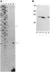
Primer extension (A) and RNase protection assays (B). Arrows indicate the presence of transcripts. A. Sequencing ladder (lane 1–4). Extension products with primer “exon II.1” (lane 5), “intron I.1” (lane 6), “exon I.2” (lane 7), and “exon I.1” (lane 8). Total RNA from C6 rat glioma cells was used in all cases. B. pBR Hga I marker numbers given in base pairs (lane m); 5 μg RNA from C6 (lane 1) and P2T (lane 2); 20 μg RNA from C6 (lane 3) and P2T (lane 4); negative control with yeast RNA (lane 5); RNA from rat Schwann cells (lane 6).
Figure 4
Figure 4
Luciferase assays. A. The transfected GDN/PN-1 promoter constructs. The p1600luc plasmid contains the promoter fragment from the internal BamH I site (b) of the first exon to the upstream EcoR I (−1545) site (e) shown in Figure 1B. The plasmid p-1600luc contains the same fragment but in the opposite orientation. The plasmid p500luc contains the promoter fragment from the BamH I (b) site upstream to the Sal I site (s; −389) indicated in Figure 1B. Open boxes represent 5′-flanking sequences, filled boxes 5′ noncoding sequences, and hatched boxes the luciferase gene. B. Luciferase activity in transiently transfected COS-7 cells. Series 1 and 2 represent two independent experiments.
Figure 5
Figure 5
Northern blot showing GDN/PN-1 mRNA expression in different cell types. C: C6 glioma cells; F: FTO2B hepatoma cells; L: L6 myoblast; P: P2T Schwannoma cells; R: Rat-1 fibroblast; and Rb: rat brain. 10 μg total RNA were used in each lane.
Figure 6
Figure 6
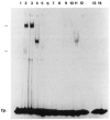
Gel shift assay of methylated and unmethylated oligonucleotides (see Materials and Methods) from the GDN/PN-1 promoter. Fractionated nuclear extracts from rat liver were incubated with methylated (lanes 1–7) and unmethylated DNA (lanes 8–14). Fractionated nuclear extract was added as follows: no extract (lanes 1 and 8), 0.1 M KCl (lanes 2 and 9), 0.2 M KCl (lanes 3 and 10), 0.3 M KCl (lanes 4 and 11), 0.4 M KCl (lanes 5 and 12), 0.5 M KCl (lanes 6 and 13), 0.6 to 1 M KCl (lanes 7 and 14). Arrows indicate the presence of methylation-specific mobility shifts, fp. the free probe.

References

    1. Adams R. L. P., Bryans M., Rinaldi A., and Yesufu H. M. I. (1990), Philos Trans R Soc Lond Biol 326, 189–198. - PubMed
    1. Artelt P., Grannemann R., Stocking C., Friel J., Bartsch J., and Hauser H. (1991), Gene 99, 249–254. - PubMed
    1. Beato M. (1989), Cell 56, 335–344. - PubMed
    1. Bird A.P. (1986), Nature 321, 209–213. - PubMed
    1. Boyes J. and Bird A. P. (1991), Cell 64, 1123–1134. - PubMed

Publication types

MeSH terms

LinkOut - more resources