. 1993;3(2):215-25.

The two functionally distinct amino termini of chicken c-ets-1 products arise from alternative promoter usage

Affiliations

The two functionally distinct amino termini of chicken c-ets-1 products arise from alternative promoter usage

P Crepieux et al. Gene Expr. 1993.

Abstract

The chicken c-ets-1 locus gives rise to two distinct transcription factors differing by structurally and functionally unrelated N-termini. p54c-ets-1 shows a striking phylogenetic conservation from Xenopus to humans, while p68c-ets-1, the cellular counterpart of the E26-derived v-ets oncogene, is apparently restricted to avian and reptilian species. In the chick embryo, both mRNAs are expressed in a wide array of tissues of mesodermal origin; however, in the embryo and after hatching, p68c-ets-1 is excluded from lymphoid cells where p54c-ets-1 accumulates. In this report, we define the basis of the differential expression of the chicken c-ets-1 products to assess their different potentials as transcription factors. We demonstrate that the two distinct N-termini arise from alternative promoter usage within the chicken c-ets-1 locus. Examination of both promoters reveals that transcription initiates from multiple sites, consistent with the absence of TATA and CAAT elements. Of these two regulatory regions, only the one that initiates the p54c-ets-1 mRNA synthesis is of the G + C-rich type, and its organization is conserved in humans. The avian-specific p68c-ets-1 promoter activity was enhanced by its own product. In addition, we identify numerous potential binding sites for lymphoid-specific transcription factors that might contribute to a tight repressor effect in lymphoid tissues.


Figures

Figure 1
Figure 1
Genomic organization of the chicken c-ets-1 locus. Open boxes mark the common exons, namely a to F; the alternative exons are indicated by striped (α and β) or dashed (I54) boxes, and the non-coding sequences are boxed in black. The two types of mRNA, which differ in the 5′ part of the gene, are shown. The Y exon and the localization of the promoters are described in this paper.
Figure 2
Figure 2
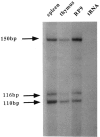
Mapping of the p54c-ets-1 transcription start sites by primer extension analysis on total RNAs extracted from chicken spleen, thymus, or the RP9 B cell line. No signal is detected in the yeast tRNA control lane. The three major start sites map 241, 207, and 201 bp upstream from the I54 initiation codon.
Figure 3
Figure 3
Nucleotide sequence of the 732 bp Hind III–Cla I DNA fragment. The mRNA major cap sites are indicated by vertical arrows, while the p54c-ets-1 cDNA 5′ end is noted with an asterisk. The G+A stretch is underlined with dashes, and the translation initiation codon of p54c-ets-1 is boxed. The position of primer 2 is indicated by a horizontal arrow. Putative recognition sequences for transcription factors are boxed. The bases are numbered with respect to the most upstream start site.
Figure 4
Figure 4
Genomic organization of the 5′ part of the chicken c-ets-1 locus. Relevant restriction sites are indicated as follows: B = BamH I; H = Hind III; P = Pst I; S = Ssp I. The Y and α exons are marked by boxes; the coding part of the α exon is striped. A double-headed arrow covers the 1204 bp DNA fragment, which was cloned into the pLUCDSS vector for testing promoter activity (fragment −1123). The three most 5′ exons included in the p68c-ets-1 cDNA are shown below. At the bottom, the sequence of the Y and α exons splice junctions is shown. The intron 5′ and 3′ boundaries are noted in lowercase letters. The conserved gt and ag nucleotides are underlined, and the initiation codon is boxed.
Figure 5
Figure 5
A. Mapping of the p68c-ets-1 transcripts start sites by RNase protection analysis on total RNA extracted from chicken spleen, E6 embryonic dermis, or thymus. Five start sites extend the Y exon beyond the Hind III restriction site of 77 to 89 bp, as indicated by arrows. B. Nucleotide sequence of the 1.5 kbp chicken genomic DNA spanning the first exon and the 5′ upstream region of the p68c-ets-1 mRNA. It is numbered with respect to the major start site. The nomenclature is the same as in Figure 3, except that the horizontal arrow marks primer 1. The Inr region is underlined. The open triangles indicate the 5′ endpoints of the deletions tested in the functional assay (see Table 1).
Figure 6
Figure 6
Effect of the p68c-ets-1 protein on its own promoter assayed in CEF. The −1123 deletion mutant sub-cloned into the pLUCDSS was transfected alone (lane 1), or with 0.5 or 1 μg of pSG5p68c-ets-1 expression vector (lanes 2 and 3, respectively). Lanes 4 and 5 show the lack of effect of the expression vector on pLUCDSS activity.

References

    1. Albagli O., Flourens A., Crépieux P., Bègue A., Stéhelin D., and Leprince D. (1992), Oncogene 7, 1435–1439. - PubMed
    1. Allen J. M., Forbush K. A., and Perlmutter R. M. (1992), Mol Cell Biol 12, 2758–2768. - PMC - PubMed
    1. Anderson S. J., Chou H. S., and Loh D. Y. (1988), Proc Natl Acad Sci USA 85, 3551–3554 - PMC - PubMed
    1. Biggin M. D. and Tjian R. (1988), Cell 53, 699–711. - PubMed
    1. Dalton S. and Treisman R. (1992), Cell 68, 597–612. - PubMed

Publication types

LinkOut - more resources