

Review
doi: 10.1165/ajrcmb/8.6.582.
Role of actin polymerization in cell locomotion: molecules and models
Affiliations
- PMID: 8323743
- PMCID: PMC4655811
- DOI: 10.1165/ajrcmb/8.6.582
Item in Clipboard
Review
Role of actin polymerization in cell locomotion: molecules and models
Am J Respir Cell Mol Biol.
1993 Jun.
Abstract
Actin filaments forming at the anterior margin of a migrating cell are essential for the formation of filopodia, lamellipodia, and pseudopodia, the "feet" that the cell extends before it. These structures in turn are required for cell locomotion. Yet the molecular nature of the "nucleator" that seeds the polymerization of actin at the leading edge is unknown. Recent advances, including video microscopy of actin dynamics, discovery of proteins unique to the leading edge such as ponticulin, the Mab 2E4 antigen, and ABP 120, and novel experimental models of actin polymerization such as the actin-based movements of intracellular parasites, promise to shed light on this problem in the near future.
Figures
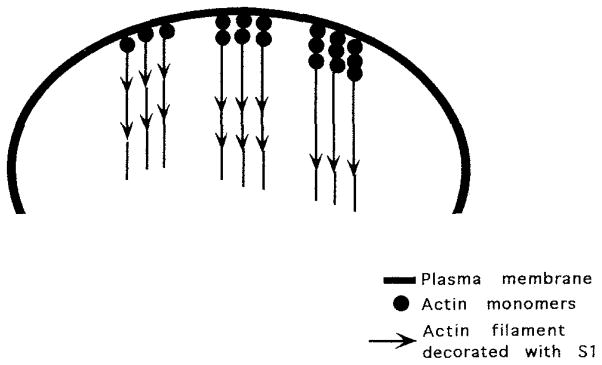
Actin filaments grow off the plasma membrane. New subunits are added to the growing filament at the membrane-filament interface. When labeled with the SI fragment of myosin, the barbed end of the filament is at the membrane, while the pointed end extends into the cell interior. Monomer must be lost from the pointed end of the filament because, after achieving a uniform length, filaments do not elongate despite the continued addition of subunits to the barbed end.
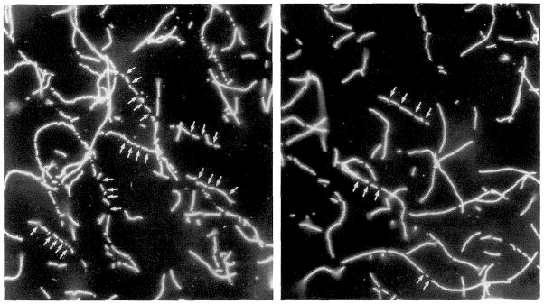
Rhodamine-phalloidin–stabilized filaments mixed with proteins eluting with ATP from filamentous actin columns are rapidly severed. These events can be visualized by fluorescence microscopy. The small arrows indicate sites where a long filament has been cut. (Reproduced by permission, J. Cell Biol. 115:1629–1638, 1991.)
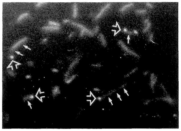
Actin filaments bind by one end to coverslips coated with capping proteins, such as gCap 39, Cap Z, or the Mab 2E4 antigen. The bound end appears bright in the micrograph (large arrow), while the dangling tail of the filament is often out of focus (small arrows) because it is hanging below the focal plane. Sometimes only the attached end is visible in the micrograph because the unattached length of filament undergoes rapid Brownian motion and does not remain in place long enough to expose the film. (Reproduced by permission, J. Cell Biol. 115:1629–1638, 1991).
References
-
- Wegner A. Head to tail polymerization of actin. J Mol Biol. 1976;108:139–150. - PubMed
Publication types
MeSH terms
Substances
Grants and funding
LinkOut - more resources
Full Text Sources
