Comparative Study
. 1993 Jul 9;616(1-2):99-104.
doi: 10.1016/0006-8993(93)90197-u.

Identification of a 34 kDa protein specific to synaptic vesicles

Affiliations
Comparative Study

Identification of a 34 kDa protein specific to synaptic vesicles

K G Miller et al. Brain Res. .

Abstract

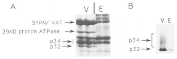
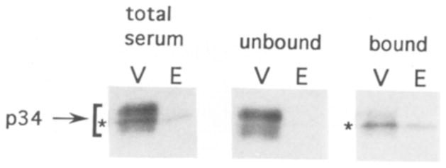
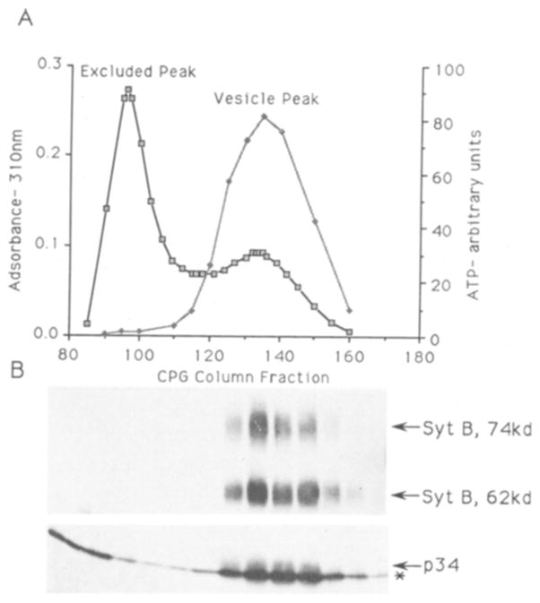
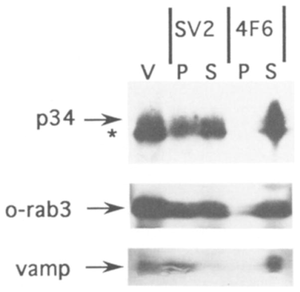
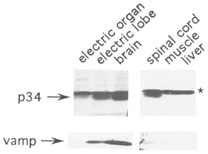
In this study, we used synaptic vesicles purified from the electric organ of marine electric rays to search for novel molecules which have important functions in synaptic transmission. Proteins that copurified with synaptic vesicles were used to immunize rats, and the resulting antisera were then used to further characterize the vesicle proteins. One of the antisera recognizes a protein of 34 kDa, p34, that has several characteristics which suggest it is a synaptic vesicle specific protein: (1) it copurifies exclusively with the synaptic vesicle peak during permeation chromatography on a controlled pore glass beads column, (2) it can be immunoprecipitated with intact synaptic vesicles and (3) it is specifically localized to the nervous system. The results suggest that p34 is a synaptic vesicle specific protein with a widespread distribution in the nervous system.


Figures

Fig. 1
Fig. 1
A: region of gel containing p34. SDS-PAGE fractionated proteins (90 μg) from the synaptic vesicle peak (V) and the excluded peak (E) of the CPG column. Proteins were visualized by Coomassie blue staining. Only the region of the gel between 30 kDa and 43 kDa is shown. Locations of the molecular weight markers are indicated on the right. The locations of synaptophysin (SYPN), VAT, the 39 kDa subunit of the proton ATPase, p34, and p32 are indicated with arrows on the left. The locations of synaptophysin and of VAT have previously been determined by Western blot analysis and amino terminal sequence analysis The locations of p32 and p34 were determined by Western blot analysis. The 39 kDa proton ATPase subunit was identified by protein sequencing of two CNBr fragments (see Materials and Methods). B: p34 is found in purified vesicles, but not excluded peak protein. Western blot analysis of an equal amount of protein (3 μg) from the excluded peak and vesicle fractions of the CPG column. The blot was probed with the antiserum raised against p32. The p34 immunoreactivity found in the same serum is evident as a diffuse band above p32.
Fig. 2
Fig. 2
Antibodies specific to p34 can be separated from p32 antibodies. Western blot analysis of proteins from the vesicle peak (V, 1 μg) and excluded peak (E, 4 μg) of the CPG column. Blots were separately probed with the p32/p34 antiserum (total serum), antibodies which did not bind to the p32 affinity column (unbound), or antibodies which were eluted from the p32 affinity column after washing (bound). The position of p34 immunoreactivity in the blots is indicated with a bracket. The position of p32 immunoreactivity, which comigrated with p34 in this gel, is indicated with an asterisk.
Fig. 3
Fig. 3
A: CPG column profile of a synaptic vesicle purification. Fractions from the final stage of the synaptic vesicle purification (the controlled pore glass beads (CPG) column were analyzed for light scattering material (membranes and protein) by adsorbance at 310 nm (open boxes) and for intact vesicles by ATP content (shaded diamonds). The fractions corresponding to membranes excluded from the glass bead pores (300 nm) and those containing synaptic vesicles are indicated. B: p34 copurifies with the synaptic vesicle containing fractions. Western blot analysis of fractions from the CPG column (equal volume). After transer, the blot was cut into strips which were probed with either Synaptotagmin B antibodies or the p32/p34 antiserum. Synaptotagmin B immunoreactivity, both the 62 kDa and 74 kDa forms, and p34 immunoreactivity are each indicated by arrows, p32 immunoreactivity is indicated by an asterisk.
Fig. 4
Fig. 4
p34 can be immunoprecipitated with SV2-containing vesicles. Western blot analysis of proteins immunoprecipitated with either a monoclonal antibody to SV2 or a control monoclonal to an Aplysia antigen (4F6). Proteins from the pellet (P) and supernatant (S) of each immunoprecipitation are indicated above the lanes. A lane of purified synaptic vesicle protein (V) was also included. Blots were cut into strips and separately probed with antibodies to p32/p34, vamp, and ommata rab3 (o-rab3).
Fig. 5
Fig. 5
p34 is specific to the nervous system. Western blot analysis of SDS-solubilized proteins (30 μg each lane) from electric organ, electric lobe (brainstem), whole brain, spinal cord, muscle, and liver. Blots were cut into strips and probed with either the p32/p34 antiserum or a polyclonal antibody to vamp, an integral membrane protein of the synaptic vesicle. Immunoreactivity corresponding to p34 and vamp are indicated with arrows, and p32 immunoreactivity is indicated with an asterisk. In the gel system used for this blot, the p34 band migrated erratically and appears below the p32 band. This was probably because a shorter resolving gel was used in combination with a different percentage of acrylamide (see methods). A control lane of synaptic vesicles was used to confirm the location of p34 immunoreactivity relative to p32.

Similar articles

References

    1. Bajjalieh SM, Peterson K, Shinghal R, Scheller RH. SV2, a brain synaptic vesicle protein homologous to bacterial transporters. Science. 1992;257:1271–1273. - PubMed
    1. Buckley K, Kelly RB. Identification of a transmembrane glycoprotein specific for secretory vesicles of neural and endocrine cells. J Cell Biol. 1985;100:1284–1294. - PMC - PubMed
    1. Carlson SS, Wagner JA, Kelly RB. Purification of synaptic vesicles from elasmobranch electric organ and the use of biophysical criteria to demonstrate purity. Biochemistry. 1978;17:1188–1199. - PubMed
    1. Cowan D, Linial M, Scheller RH. Torpedo synaptophysin: evolution of a synaptic vesicle protein. Brain Res. 1990;509:1–7. - PubMed
    1. Feany MB, Lee S, Edwards RH, Buckley KM. The synaptic vesicle protein SV2 is a novel type of transmembrane transporter. Cell. 1992;70:861–867. - PubMed

Publication types

MeSH terms