Comparative Study
. 1993 Apr;8(1):143-57.
doi: 10.1111/j.1365-2958.1993.tb01211.x.

Dual roles of plcA in Listeria monocytogenes pathogenesis

Affiliations
Comparative Study

Dual roles of plcA in Listeria monocytogenes pathogenesis

A Camilli et al. Mol Microbiol. 1993 Apr.

Abstract

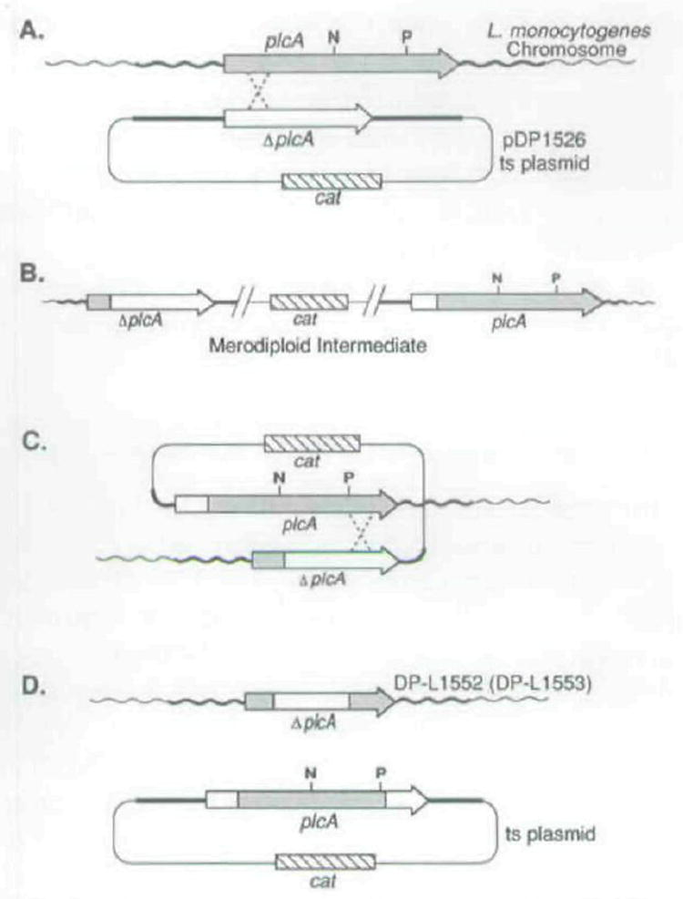
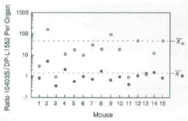
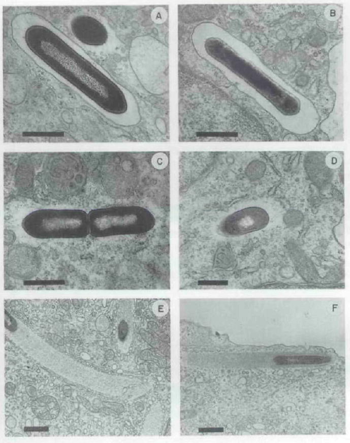
The plcA gene of Listeria monocytogenes encodes a secreted phosphatidylinositol-specific phospholipase C (Pl-PLC). Recent studies have established that transposon mutations within plcA result in avirulence for mice and pleiotropic effects when examined in tissue-culture models of infection. Genetic analysis reveals that many of the effects of the transposon insertions are due to loss of readthrough transcription from plcA into the downstream gene prfA, which encodes an essential transcription factor of numerous L. monocytogenes virulence genes. Construction of an in-frame deletion within plcA had no effect on expression of prfA thus allowing direct assignment of a role of the Pl-PLC in pathogenesis. Pl-PLC was shown to play a significant role in mediating escape of L. monocytogenes from phagosomes of primary murine macrophages. Interestingly, this defect manifested itself in vivo in the liver but not in the spleen of infected mice.


Figures

Fig. 1
Fig. 1
A Physical map of the L. monocytogenes chromosomal region containing plcA and prfA. The site of Tn917-LTV3 insertion in DP-L1054 is indicated by the arrow above the plcA gene (Sun et al., 1990). Stem and loop structures represent the locations of rho-independent terminator-like sequences (Mengaud et al., 1989; 1991). Promoters are indicated below the genes by arrows (Mengaud et al., 1989; . N, Freitag and D. A. Portnoy, unpublished). B. Complementation derivative strains of DP-L1054 and the respective L. monocytogenes DNA sequences (solid lines) contained by their comple-menting plasmids
Fig. 2
Fig. 2
Plaque formation by L. monocytogenes strains in L2 cell monolayers. Plaques were stained with neutral red after 3 d. Strains are indicated above each well. The mean plaque diameter formed by each strain is shown at the bottom of each well as a percentage of the 10403S (wild-type) mean plaque diameter. The standard deviations of ail means was ≤9% of the wild-type mean plaque diameter.
Fig. 3
Fig. 3
Upper panel. Physical map of the chromosomal region of L. monocytogenes containing the plcA and prfA genes. The directions of transcription and RNA transcripts are indicated schematically below each gene. DNA probes used in the detection of RNA transcripts are lettered A and B. Lower panel. Northern blot analysis autoradiograms of RNA transcripts from strains SLCC 5764 (lane 1), DP-L1387 (lane 2), DP-L1535 (lane 3), DP-L1553 (lane 4), 10403S (lane 5), DP-L1054 (lane 6), DP-L1510 (lane 7), and DP-L1552 (lane 8). After a 24 h exposure to generate the first autoradiogram on the left, the Northern filter membrane was washed to remove probe A and then rehybridized with probe B and exposed for 48 h to generate the second autoradiogram on the right (see the Experimental procedures). Equivalent amounts of RNA (25 μg) were analysed for each strain. The positions and approximate sizes of the 23S, 16S, and 5S ribosomal RNAs served as molecular weight markers and are indicated between the autoradiograms. The slowly migrating bands present for DP- L1054 (lane 6. probe A) represent chimaeric RNA transcripts resulting from a transcriptional fusion with Tn917-LTV3 which was inserted into the plcA gene in this strain. Similarly, the taint band of approximately 2.4 kb present for DP-L1510 (lane 7, probe A) resulted from a transcriptional fusion of plcA with one end of the integrated plasmid in this strain. A chimaeric RNA transcript of identical mobility was seen for DP-L1535 (with probe A) upon longer exposures (data not shown).
Fig. 4
Fig. 4
Schematic diagram for construction of the isogenic strains DP-L1510 and DP-L1535, in which readthrough transcription of prfA was blocked. A. Chromosomal integration of pDP1498 by homologous recombination between the cloned Pstl (P)- and EcoRI (E)-generated fragment present on the plasmid and the homologous chromosomal sequence. The designated cross-over points are arbitrary. P1 and P2 indicate the two promoter elements immediately upstream of prfA while PplcA indicates the single plcA promoter (Mengaud et al., 1989; ; N. Freitag unpublished). B. The resulting integration structure on the chromosome of both mutant strains. The integration strains were selected for, and maintained by, growth at a non-permissive temperature for plasmid replication in the presence of chloramphenicol. The plasmid was shown by Southern blot analysis to have integrated multiple times in a head-to-head configuration (data not shown).
Fig. 5
Fig. 5
Plaque formation by wild-type L. manocytogenes (10403S), a mutant strain having readthrough transcription of prfA blocked by an integrated plasmid (DP-Ll510), and two control random integration strains (DP-L1532 and DP-L1534). Plaques were stained with neutral red after 3 d. Strains are indicated above each well. The mean plaque diameter formed by each strain is shown at the bottom of each well as a percentage of the 10403S (wild-type) mean plaque diameter. The standard deviations of all means was ≤9% of the wild-type mean plaque diameter.
Fig. 6
Fig. 6
Schematic diagram for construction of the isogenic strains DP-L1552 and DP-L1553 each containing an in-frame Internal deletion in plcA. A. Chromosomal integration of pDP1526 by homologous recombination between the ΔplcA allele on the plasmid, which had the internal Nsil (N) to Pstl (P) fragment deleted, and the wild-type plcA chromosomal allele. The designated cross-over points are arbitrary; however, recombination occurred on either side of the internal deletion. B. The resulting integration structure on the chromosome which was selected for by growth at a non-permissive temperature for plasmid replication in the presence of chloramphenicol. Upon passage of the merodiploid intermediate strains for several generations without selective pressure, spontaneous excision of the integrated plasmid from the chromosome occurred (C). After curing of the excised plasmids at a non-permissive temperature for plasmid replication (D), L. monocylogenes Cms revertants were recovered at a frequency of = 1 %. Approximately 50% of the Cms revertants retained the ΔplcA allele on the chromosome. These resulted from excision of the integrated plasmid via homologous recombination on the opposite side of the deletion allele as shown in (C).
Fig. 7
Fig. 7
Coomassie-brilliant-blue stained SDS–PAGE of secreted polypeplides of mutant and wild-type L. monocytogenes SLCC 5764. Proteins were precipitated from culture supernatant fluids with 10% trichloroacetic acid and separated on a 7% SDS-polyacrylamide gel. A. Molecular mass markers of the sizes indicated (in kDa) on the left; B, parental strain SLCC 5764; C, DP-L1553; D, DP-L1387; E, DP-L1535; F, DP-L1536. Arrows indicate polypeptide species of reduced expression in DP-L1387 and DP-L1535. LLO is the major band migrating at =58 kDa indicated by the third arrow from the top (Kathariou et al., 1987). The two secreted forms of PC-PLC are indicated by the bottom and fourth from bottom arrows (Raveneau et al., 1992; Kathariou et al., 1990; D. A. Portnoy, unpublished). The second arrow from the bottom indicates a doublet consisting of PI-PLC (top band), which accordingly is absent from DP-L1553 (compare lanes B and C), and an unknown polypeptide (bottom band).
Fig. 8
Fig. 8
Ratio of wild-type L. monocytogenes to DP-L1552 in livers (open circles) and spleens (closed circles) of mice 48 h after infection. Note that the ratio ordinate is a log scale. Fifteen mice were injected intravenously with a sublethal dose of log-phase bacteria, sacrificed after 48 h, and the number of colony-forming units per organ was determined by plating serial dilutions of organ homogenates on agar media. The ratios of wild-type (PI-PLC+) to DP-L1552 (PI-PLC) were determined by patching 200 colonies (derived from each organ homogenate) onto BHI/egg yolk agar and scoring for an opaque zone surrounding each PI-PLC+ isolate, after 24 h of bacteria growth (see the Experimental procedures). The mean ratios in liver and spleen are indicated on the right side of the figure. The ratio values for liver and spleen were statistically analysed for deviation from a value of 1 by the Wilcoxon paired sample test (Zar, 1974).
Fig. 9
Fig. 9
Intracellular growth of L. monocytogenes strains in primary and established murine macrophages cells. A, J774 cell-line; B, peritoneal-derived macrophages; C, bone marrow-derived macrophages, ○, wild-type strain 10403S; ●, DP-L1552; □, DP-L1054: ■, DPL1044. Data points and error bars represent the mean and standard deviations of the number of viable bacteria recovered from three coverslips.
Fig. 10
Fig. 10
Representative electron micrographs of thin sections of portions of bone marrow-derived macrophages infected with stationary-phase L-monocytogenes and fixed in situ. A, C. E, wild-type strain 10403S after 1, 1.5, and 4 h of infection, respectively; B, D, F, DP-L1552 alter 1, 1.5, and 4 h of infection, respectively. The presence of host membrane surrounding bacteria is shown in (A) and (B). while bacteria free in the cytoplasm are shown in the remaining panels. Actin tails were seen trailing both the wild-type and mutant L. monocytogenes bacteria after 4 h of infection (panels E and F). It is worth noting the presence in panel E, and to a lesser extent in panel F, of many small vesicles ‘coating’ the periphery of the actin tails. This was observed infrequently in electron micrographs from this experiment as well as from other experiments using different host cell types (data not shown), and thus may be highly dependent upon variable fixation conditions. The function and/or significance of these small vesicles is not known. Bars = 0.1 μm.

References

    1. Behnke D, Gilmore MS. Location of antibiotic resistance determinants, copy control, and replication functions on the double-selective streptococcal cloning vector pGB301. Mot Gen Genet. 1981;184:115–120. - PubMed
    1. Breton CB, Langsley G, Barale J, Pereira da Silva LH. A malaria phosphatidylinositol-specific phospholipase C: a possible role in merozoite maturation and erythrocyte invasion. In: Cohen CM, Palek J, editors. Cellular and Molecular Biology of Normal and Abnormal Erythroid Membranes. New York: Wiley-Liss; 1990. pp. 315–332.
    1. Camilli A, Portnoy DA, Youngman P. Insertional mutagenesis of Listeria monocytogenes with a novel Tn917 derivative that allows direct cloning of DNA flanking transposon insertions. J Bacteriol. 1990;172:3738–3744. - PMC - PubMed
    1. Camilli A, Goldfine H, Portnoy DA. Listeria monocytogenes mutants lacking phosphatidylinositol-specific phospholipase C are avirulent. J Exp Med. 1991;173:751–754. - PMC - PubMed
    1. Chakraborty T, Leimeister-Wachter M, Domann E, Hartl M, Goebel W, Nichterlein T, Notermans S. Coordinate regulation of virulence genes in Listeria monocytogenes requires the product of the prfA gene. J Bacteriol. 1992;174:568–574. - PMC - PubMed

Publication types

MeSH terms

Substances

LinkOut - more resources