. 1993 Jul;44(1):173-9.

Differential down- and up-regulation of rat brain opioid receptor types and subtypes by buprenorphine

Affiliations

Differential down- and up-regulation of rat brain opioid receptor types and subtypes by buprenorphine

M M Belcheva et al. Mol Pharmacol. 1993 Jul.

Abstract

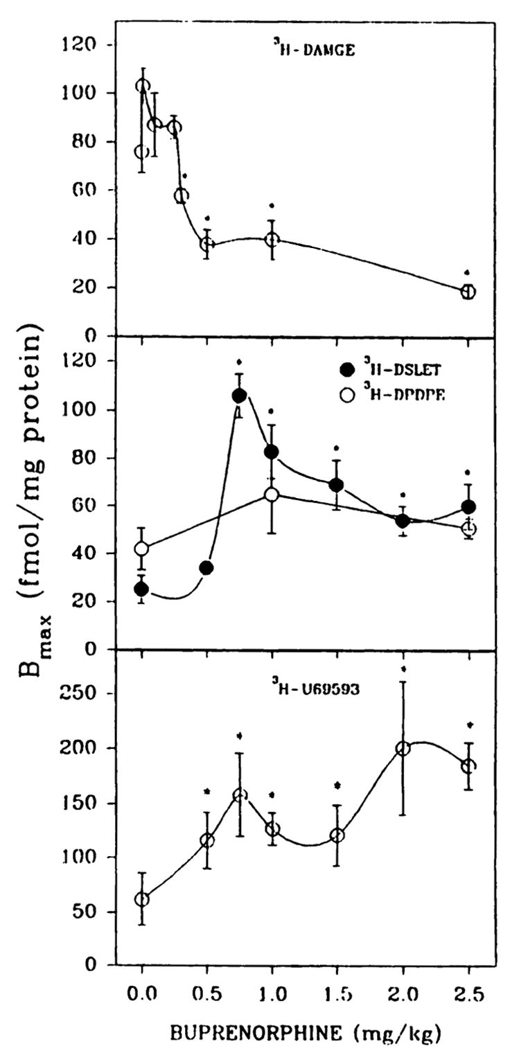
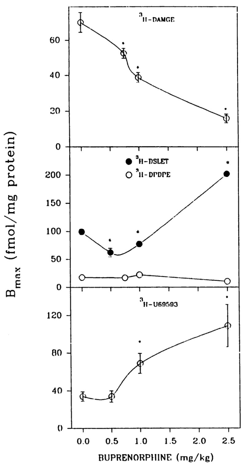
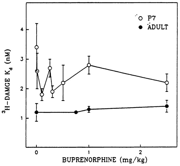
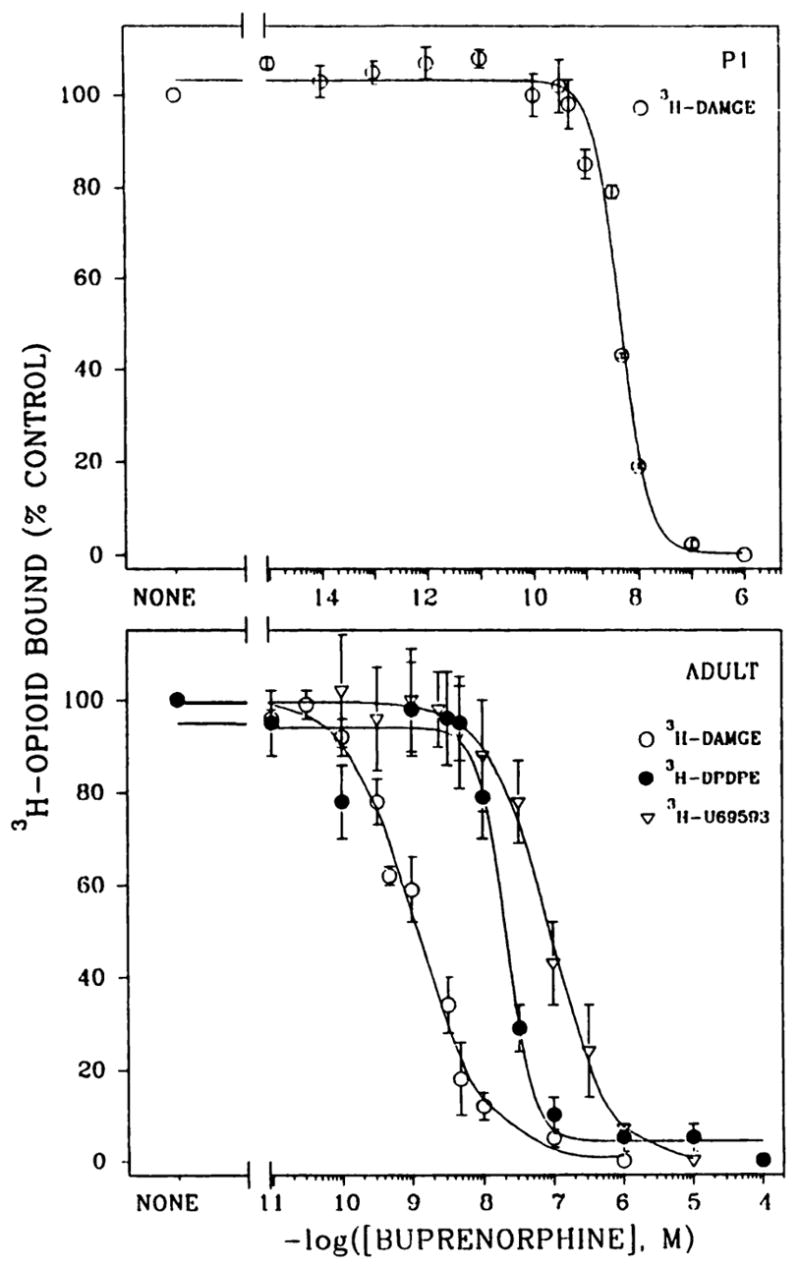
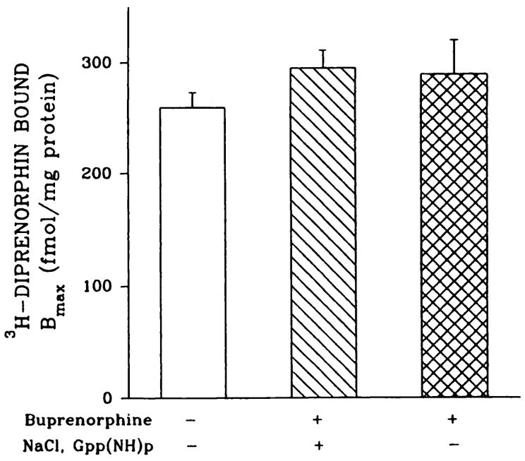
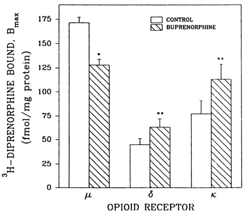
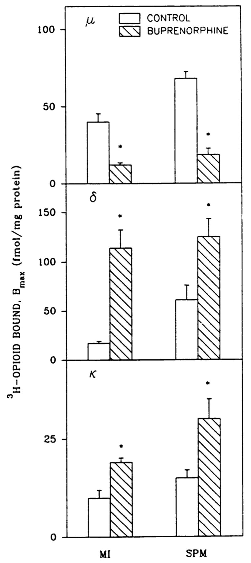
The induction of opioid receptor adaptation by mixed agonist-antagonists such as buprenorphine has not been investigated. To this end, neonatal rats were given injections of buprenorphine (0.1-2.5 mg/kg/day) and mu binding (Kd and Bmax) to brain membranes was measured with [3H][D-Ala2,MePhe4,Gly-ol5]enkephalin. At doses of buprenorphine of > or = 0.5 mg/kg, mu sites were reduced 47-75%, without changes in affinity. Chronic administration of the structurally related partial agonist diprenorphine (2.5-75 mg/kg) failed to alter mu binding. Apparent loss of sites due to receptor blockade by residual buprenorphine was ruled out by several lines of evidence. Bmax values for delta ([3H][D-Ser2,L-Leu5]enkephalyl-Thr) and kappa ([3H]U69593) binding were elevated 1.9-4.2-fold by buprenorphine treatment. In adult rats buprenorphine (0.5-2.5 mg/kg) reduced mu-opioid binding to forebrain membranes dose dependently, by 25-77%. [3H][D-Ser2,L-Leu5] Enkephalyl-Thr-labeled delta subtype receptors and kappa sites in adult forebrain membranes were up-regulated 2-3-fold. The delta subtype receptors that bind [3H][D-Pen2,D-Pen5]enkephalin in neonatal or adult brain membranes were unaffected by 0.5-2.5 mg/kg buprenorphine treatment. Down-regulation (70-74%) of mu sites and up-regulation (1.9-6.7 fold) of delta and kappa receptors were also observed in synaptic plasma membrane-enriched and microsomal fractions from buprenorphine-treated adult rat brain. Because agonist-induced opioid receptor down-regulation is difficult to elicit in adult mammalian brain, these data indicate that buprenorphine is a useful tool to study brain opioid receptor adaptation in vivo.


Figures

Fig. 1
Fig. 1
Opioid binding to brain membrane preparations from P7 rats treated in vivo with various concentrations of buprenorphine. Pups were given subcutaneous injections of saline or buprenorphine daily for 6 days and were sacrificed 20 hr after the last administration. Brain membranes were washed five times with 25–30 ml of 50 mM Tris·HCl, pH 7.4, before binding assays. Opioid binding was measured with 1 nM [3H]DAMGE, [3H]DSLET, or [3H]U69593 or 2 nM [3H]DPDPE in 12-point homologous competition assays. Bmax, and Kd values were estimated using the LIGAND program. The affinity of [3H]DAMGE (μ) is shown in Fig. 3. In the order of increasing buprenorphine concentrations from 0 to 2.5 mg/kg, Kd values were 3.3 ± 0.6, 3.3 ± 0.2, 7.6 ± 1.2, 6.2 ± 1.1, 2.8 ± 0.2, 6.0 ± 0.9, and 5.1 ± 0.7 nM ([3H]DSLET); 3.8 ± 0.6, 5.3 ± 1.3, and 3.4 ± 0.2 nM ([3H]DPDPE); and 3.1 ± 0.7, 5.1 ± 0.9, 4.2 ± 0.8, 6.4 ± 0.9, 4.9 ± 1.2, 5.8 ± 1.4, and 4.9 ± 0.3 nM ([3H]U69593). *, Significantly different from saline treated, p < 0.05. Results are from four to seven separate experiments.
Fig. 2
Fig. 2
Optoid binding in brain membrane preparations from adult rats treated in vivo with various concentrations of buprenorphine. Adult male rats were administered (intraperitoneally) either buprenorphine (0.5–2.5 mg/kg) or saline, and 20 hr later forebrains were collected. The affinity of [3H]DAMGE (μ) is shown in Fig. 3. In the order of increasing bupren­orphine concentrations from 0 to 2.5 mg/kg, Kd values were 2.2 ± 0.2, 2.0 ± 0.3, 2.2 ± 0.3, and 4.6 ± 0.6 nM ([3H]DSLET); 2.0 ± 0.2, 2.0 ± 0.3, 2.4 ± 0.2, and 1.6 ± 0.3 nM ([3H]DPDPE); and 3.3 ± 0.6,6.2 ± 1.2, 4.3 ± 0.5, and 5.7 ± 1.5 nM ([3H]U69593. *, Significantly different from saline-treated, p < 0.05. Results are from three to six separate experiments.
Fig. 3
Fig. 3
Ka values for [3H]DAMGE binding to membrane preparations from P7 and adult rats treated with buprenorphine. Rats were given injections of buprenorphine as described for Figs. 1 and 2. [3H]DAMGE Kd estimations were obtained from 12-point homologous competition binding assays. Note that P7 control Ka values were consistently higher than the Kd values for buprenorphine-treated rats. Results are from six or seven separate experiments.
Fig. 4
Fig. 4
Heterologous competition binding curves with buprenorphine as competitor for μ, δ, and κ receptor sites. [3H]DAMGE (1 nM) binding was determined in P1 (upper) and adult (lower) brain membranes in the presence of 12–16 concentrations of buprenorphine. [3H]DPDPE (2 nM) and [3H]U69593 (1 nM) binding was conducted with adult brain membranes. Corresponding Bmax and Ki values are given in Table 1. Results are from three to nine separate experiments.
Fig. 5
Fig. 5
Optoid binding to NaCl/Gpp(NH)p-washed brain membranes from buprenorphine-treated adult rats. Adult rats were given injections of 0.5 mg/kg buprenorphine. Forebrains were divided into two hemispheres; one, used for membrane preparations, was washed with 50 mM Tris·HCl, pH 7.4, buffer and the other was washed with 50 mM Tris·HCl, pH 7.4, buffer containing 100 mM NaCl and 50 μM Gpp(NH)p, followed by four additional washes with buffer alone. Homologous displacement assays were performed with 1 nM [3H]diprenorphine and Bmax values were determined with the INPLOT4 program. Kd values varied from 1.1 ± 0.1 to 4.4 ± 0.9 nM. Results are from four or five separate experiments.
Fig. 6
Fig. 6
[3H]Diprenorphine binding to brain membranes from buprenorphine-treated adult rats. Animals were given one intraperitoneal injection of 0.5 mg/kg buprenorphine 20 hr before sacrifice. Membranes were incubated for 2 hr with 50 mM Tris·HCl, pH 7.4, buffer containing 100 mm NaCl and 50 μM Gpp(NH)p, followed by four additional washes with buffer alone. Opioid binding was measured with 1 or 6 nM [3H]diprenorphine, and unlabeled diprenorphine (5 μM) was used to determine non-specific binding, as described in Materials and Methods. *, p < 0.05; **, p < 0.01. Results are from three separate experiments.
Fig. 7
Fig. 7
Down- and up-regulation of opioid receptors in subcellular fractions from brain of buprenorphine-treated adults. SPMs and microsomes (MI) from adult rats treated with saline or buprenorphine (1 mg/kg, 20 hr) were prepared as described in Materials and Methods. Bmax values were estimated with 1 nM [3H]DAMGE (μ), [3H]DSLET (δ), and [3H] U69593 (κ) in homologous competition assays. For microsomes and SPMs, control Kd values were 1.8 ± 0.1 and 1.4 ± 0.07 nM (μ), 1.3 ± 0.2 and 3.3 ± 0.6 nM (δ), and 1.9 ± 0.4 and 1.2 ± 0.1 nM (κ), whereas those for buprenorphine-treated membranes were 5.5 ± 2.5 and 1.9 ± 0.3 nM (μ), 3.7 ± 0.5 and 3.9 ± 0.5 nM (δ), and 3.5 ± 0.1 and 3.3 ± 0.4 nM (κ), respectively. *, Significantly different from saline-treated rats, p < 0.05. Results are from three to five separate experiments.

References

    1. Mello NK, Mendelson JH. Buprenorphine suppresses heroin use by heroin addicts. Science (Washington D C) 1980;207:657–659. - PubMed
    1. Kosten TR, Kleber HD, Morgan C. Treatment of cocaine abuse with buprenorphine. Biol Psychiatry. 1989;26:637–639. - PubMed
    1. Johnson RE, Jaffe JH, Fudala PJ. A controlled trial of buprenorphine treatment for opioid dependence. JAMA. 1992;267:2750–2755. - PubMed
    1. Cowan A, Lewis JW, MacFarlane IR. Agonist and antagonist properties of buprenorphine, a new antinociceptive agent. Br J Pharmacol. 1977;60:537–545. - PMC - PubMed
    1. Jasinski DR, Pevnick JS, Griffith D. Human pharmacology and abuse potential of the analgesic buprenorphine. Arch Gen Psychiatry. 1978;35:601–616. - PubMed

Publication types

LinkOut - more resources