

Molecular analysis of transforming growth factor beta in giant cell tumor of bone
- PMID: 8500096
- PMCID: PMC5474756
- DOI: 10.1016/0165-4608(93)90237-g
Molecular analysis of transforming growth factor beta in giant cell tumor of bone
Abstract
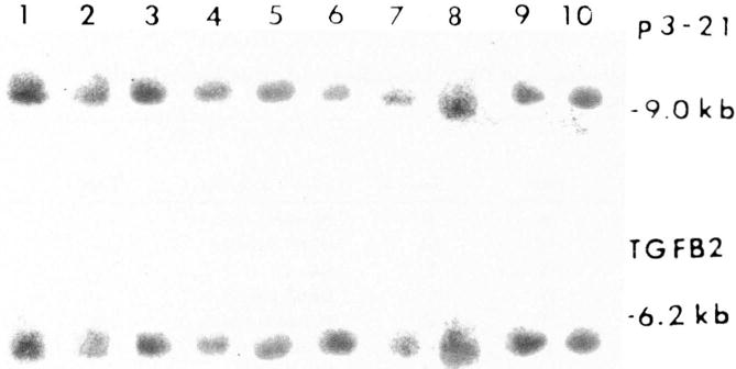
Giant cell tumor of bone (GCT) is a primary bone neoplasm with unique cytogenetic findings including telomeric associations. Elevated expression of message RNA for transforming growth factor beta (TGF beta), but not transforming growth factor alpha (TGF alpha), has been reported in this tumor. Further investigation of GCT was undertaken to determine whether genetic loci for TGF beta in GCT patients with and without chromosome abnormalities are altered. Due to the reported TGF beta overexpression in GCT, qualitative and quantitative Southern blot analyses with TGF beta 1 and TGF beta 2 and an internal control probe (p3-21) were performed with tumor DNA and DNA from normal tissue on ten patients with GCT and control individuals. No obvious TGF beta 1 or TGF beta 2 gene alterations were detected. Normal copy numbers were calculated when comparing tumor and normal DNA from GCT patients as well as DNA from control individuals. Abnormal chromosome findings, including telomeric associations, marker chromosome, double minutes, chromosome fragments, ring chromosomes (possibly representing intra-chromosome telomeric associations), and polyploid cells were observed in seven of the ten patients with GCT. Chromosomes 11, 16, 19, 20, and 21 were most commonly observed in telomeric associations, with the terminus of the long arm of chromosome 19 being the most frequent. We conclude that there are no TGF beta 1 or TGF beta 2 gene alterations detected in GCT with the methodologies described, and that telomeric associations are a reproducible cytogenetic characteristic of this neoplasm.
Figures


References
-
- Schwartz HS, Jenkins RB, Dahl RJ, Dewald GW. Cytogenetic analyses on giant cell tumors of bone. Clin Orthop Rel Res. 1989;240:250–260. - PubMed
-
- Bridge JA, Neff JR, Bhatia PS, Sanger WG, Murphey MR. Cytogenetic findings and biologic behavior of giant cell tumors of bone. Cancer. 1990;65:2697–2703. - PubMed
-
- Bridge JA, Neff JR, Mouron BJ. Giant cell tumor of bone: Chromosomal analysis of 48 specimens and review of the literature. Cancer Genet Cytogenet. 1992;58:2–15. - PubMed
-
- Bardi G, Pandis N, Mandahl N, Helm S, Sfikas K, Willen H, Panagiotopoulos G, Rydhold A, Mitelman F. Chromosomal abnormalities in giant cell tumors of bone. Cancer Genet Cytogenet. 1991;57:161–167. - PubMed
Publication types
MeSH terms
Substances
Grants and funding
LinkOut - more resources
Full Text Sources
Medical
