. 1993;3(1):95-107.

p53 domains: suppression, transformation, and transactivation

Affiliations

p53 domains: suppression, transformation, and transactivation

M Reed et al. Gene Expr. 1993.

Abstract

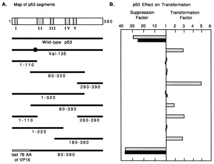
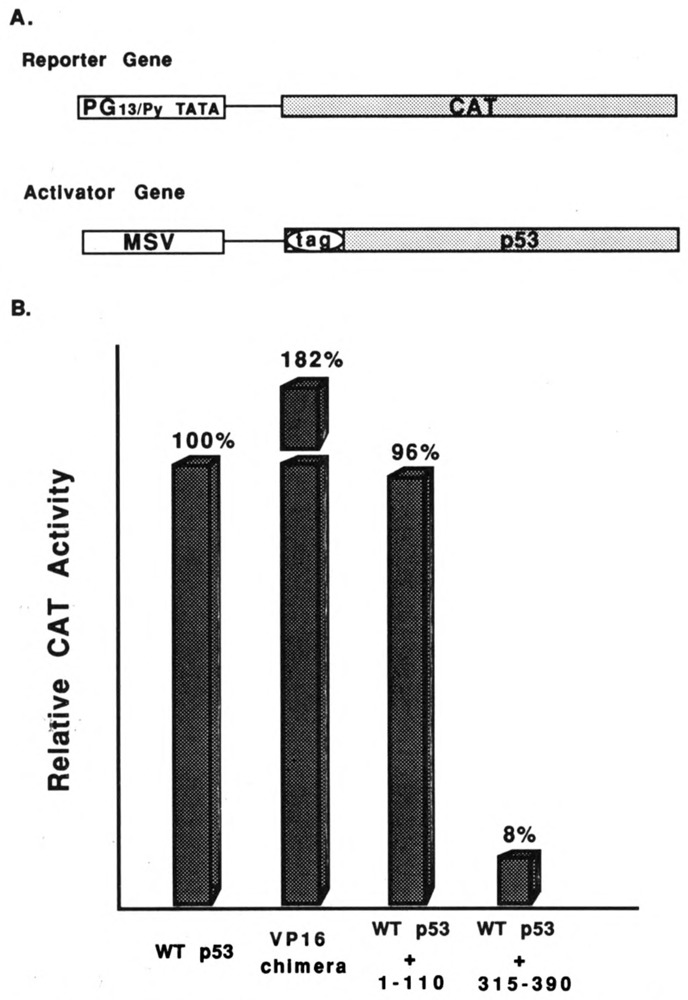
We investigated the suppression, transformation, and transactivation functions of isolated segments of wild-type murine p53. Intact p53, but no segment of p53, inhibited cellular transformation by the activated ras and adenovirus E1A proteins. We conclude that most of p53 is needed for suppression of cellular proliferation. Nevertheless, the transactivating domain of herpesvirus protein VP16 was able to substitute for the N-terminal transactivating domain of p53 in cellular suppression. Thus, unless the interchanged p53 and VP16 acidic segments share additional functions, transactivation is required for suppression by p53. Interestingly, we found that all p53 segments containing amino acids 320-360 enhanced transformation by ras and E1A. This region has been associated with the oligomerization of p53 (Milner et al., 1991; Sturzbecher et al., 1992). Furthermore, no p53 segment lacking amino acids 320-360 transformed cells. Amino acids 320-360, therefore, may account for the major transforming activity of p53. Intact p53 and chimeric VP16-p53 transactivated the CAT gene under control of a p53-specific promoter, while transforming segments of p53 interfered with transactivation by wild-type p53. Our findings argue that transactivation by p53 is required for cellular suppression and that any nontransactivating p53 that retains the capacity to oligomerize with wild-type p53 would have transformation potential.


Figures

Figure 1
Figure 1
Vector for the expression of p53 and domains of p53. The vector contains the MSV promoter, a cassette for insertion of protein segments, an RNA splice signal, and a polyadenylation signal. Insertion of coding sequences in-frame in the cassette creates a tagged protein. The tag contains an optional E1A nuclear localization signal (NLS), six histidines for metal affinity purification, the KT3 epitope, and a site for cleavage by proteinase Xa.
Figure 2
Figure 2
Suppression and transformation by murine p53 and domains of p53. A. Structures of the p53 polypeptides. WT p53 has five regions of conservation; mutations in regions II–V are frequent sites of mutations in human tumors and in transformed mouse cells. The solid lines represent p53 segments expressed by different plasmids. B. Suppression and transformation activities of the polypeptides shown in A. Test plasmids expressing no p53, wild-type p53, or isolated segments of p53 were cotransfected with plasmids expressing activated ras and E1A into REF cells. Transformed foci were counted 12 days after transfection. The levels of suppressed or enhanced transformation were determined by comparing the number of foci induced by p53 segments, ras, and E1A to the number of foci induced by ras and E1A, as described in the text. Shaded bars indicate p53s without an E1A NLS; solid bars indicate p53s with an E1A NLS added to their N-terminal tags.
Figure 3
Figure 3
Examples of transformed foci induced by selected plasmids shown in Figure 2. Test plasmids, identified in the figure, were cotransfected into REF cells. Transformed foci were stained with Coomassie Blue 12 days after transfection.
Figure 4
Figure 4
Fine mapping of the transformation domain of p53. Test plasmids expressing no p53, p53Val135, or isolated segments of p53 were cotransfected with plasmids expressing ras and E1A or ras alone into REF cells. The total DNA added to all cultures was 10 μg. Transformed foci were counted 12 days after transfection. We quantitated the transforming potential of the truncated polypeptides by measuring their enhancement of transformation by ras and E1A or by ras alone in the absence (shaded bars) or in the presence (solid bars) of an artificial nuclear localization signal.
Figure 5
Figure 5
Transactivation by wild-type and mutant p53s. HCT-116 cells were transfected with plasmids expressing the CAT gene under the control of the p53-specific PG13/Py promoter and wild-type p53 or segments of p53. A. General structure of the CAT reporter gene and the genes expressing wild-type p53 or segments of p53. B. Levels of CAT activity induced by the various p53s or combinations of p53 indicated at the bottom of the histogram.
Figure 6
Figure 6
Expression of tagged p53 in REF and SF9 cells. A. REF cells were transfected with pAT plasmids expressing wild-type p53 or segments of p53. The tagged p53s were concentrated by immunoprecipitation and analyzed by immunoblotting with KT3 monoclonal antibodies, as described in Materials and Methods. B. SF9 cells were infected with baculovirus expressing wild-type p53 or segments of p53. Tagged p53s were identified directly by immunoblotting with KT3 monoclonal antibodies, as described in Materials and Methods. The N + C abbreviation designates the p53 segment containing N-terminal (1–110) and C terminal (280–390) segments of p53.
Figure 7
Figure 7
Genetic and structural organization of p53. The protein is divided into three regions: an N-terminal acidic transactivation domain, a central domain with conserved sequences and highly specialized functions, and a C-terminal oligomerization domain. The optimal transformation domain coincides with the oligomerization domain. The sequences of the core transforming domain from 315–360 are shown and compared with similar sequences of p53s from other species. The shaded sequences are the most conserved.

References

    1. Aoyama N., Nagase T., Sawazaki T., Mizuguchi G., Nakagoshi H., Fujisawa J., Yoshida M., and Ishii S. (1992), Proc Natl Acad Sci USA 89, 5403–5407. - PMC - PubMed
    1. Baker S. J., Fearon E. R., Nigro J. M., Hamilton S. R., Preisinger A. D., Jessup J. M., vanTuinen P., Ledbetter D. H., Barker D. F., Nakamura Y., White R., and Vogelstein B. (1989), Science 244, 217–221. - PubMed
    1. Baker S. J., Markowitz S., Fearon E. R., Willson J. K. V., and Vogelstein B. (1990), Science 249, 912–915. - PubMed
    1. Barak Y. and Oren M. (1992), EMBO J 11, 2115–2121. - PMC - PubMed
    1. Bargonetti J., Friedman P. N., Kern S. E., Vogelstein B., and Prives C. (1991), Cell 65, 1083–1091. - PubMed

Publication types